. 1999 Nov 23;96(24):13679-84.
doi: 10.1073/pnas.96.24.13679.

Antibody C219 recognizes an alpha-helical epitope on P-glycoprotein

Affiliations

Antibody C219 recognizes an alpha-helical epitope on P-glycoprotein

J M van Den Elsen et al. Proc Natl Acad Sci U S A. .

Abstract

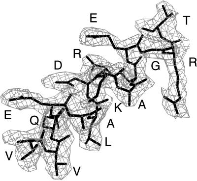
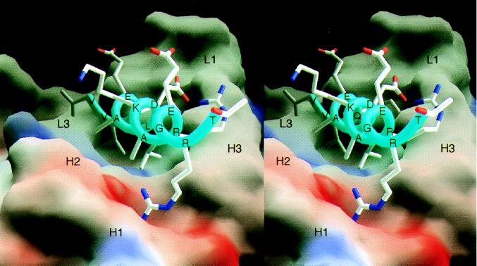
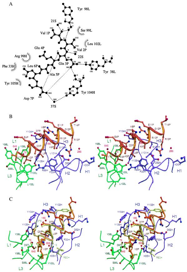
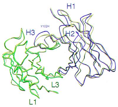
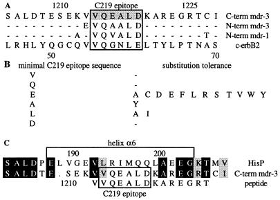
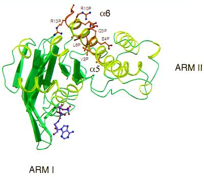
The ABC transporter, P-glycoprotein, is an integral membrane protein that mediates the ATP-driven efflux of drugs from multidrug-resistant cancer and HIV-infected cells. Anti-P-glycoprotein antibody C219 binds to both of the ATP-binding regions of P-glycoprotein and has been shown to inhibit its ATPase activity and drug binding capacity. C219 has been widely used in a clinical setting as a tumor marker, but recent observations of cross-reactivity with other proteins, including the c-erbB2 protein in breast cancer cells, impose potential limitations in detecting P-glycoprotein. We have determined the crystal structure at a resolution of 2.4 A of the variable fragment of C219 in complex with an epitope peptide derived from the nucleotide binding domain of P-glycoprotein. The 14-residue peptide adopts an amphipathic alpha-helical conformation, a secondary structure not previously observed in structures of antibody-peptide complexes. Together with available biochemical data, the crystal structure of the C219-peptide complex indicates the molecular basis of the cross-reactivity of C219 with non-multidrug resistance-associated proteins. Alignment of the C219 epitope with the recent crystal structure of the ATP-binding subunit of histidine permease suggests a structural basis for the inhibition of the ATP and drug binding capacity of P-glycoprotein by C219. The results provide a rationale for the development of C219 mutants with improved specificity and affinity that could be useful in antibody-based P-glycoprotein detection and therapy in multidrug resistant cancers.


Figures

Figure 1
Figure 1
Electron density corresponding to the helical peptide epitope in molecule II. Density is calculated with coefficients |Fobs − Fcalc|ph(calc) before incorporation of the peptide into the model. The final conformation of the epitope is superimposed in thick bonds. Residues in the epitope are identified by single letter residue type. This figure was prepared with the program o (28).
Figure 2
Figure 2
Molecular surface representation of the scFv C219 (molecule II) binding site with the bound α-helical P-glycoprotein epitope peptide. The molecular surface is colored for electrostatic potential (red for negative charge, blue for positive charge). Peptide residues and the approximate locations of C219 heavy (H) and light chain (L) hypervariable loops are indicated. Fig. 2 was produced with the program grasp (32).
Figure 3
Figure 3
Interactions between the α-helical peptide and the C219 binding site. (A) Two-dimensional ligplot (33) representation of the interactions between residues of the minimal NBD-epitope peptide (P), C219 heavy (H) and light chain (L) residues, and solvent molecules (S), as seen in molecule I. The residues that form van der Waals contacts with the peptide are depicted as labeled arcs with radial spokes pointing toward the peptide atoms with which they interact. C219 residues that form hydrogen bonds are shown in a ball-and-stick representation, and the hydrogen bonds are presented as dashed lines. Of all of the intrapeptide hydrogen bonds present in the structure, only the bonds between Gln 3P and Asp 7P are shown. (B) Stereoplot of the Fv-peptide interactions seen in molecule II. (C) Comparison of the bound NBD-epitope peptide in molecule I and II. In B and C, light (L) and heavy chain (H) residues and backbone positions of the scFv C219 are shown in green and magenta. Peptide backbone and side chains are shown in khaki for molecule I and in gold for molecule II. Positions of water molecules are indicated as red spheres. Different positions of binding site residues and water molecules in molecule I are also colored khaki. B and C were generated by using molscript (34) and raster3d (35).
Figure 4
Figure 4
Relative positions of the variable light and heavy chains in the dimers of the unliganded scFv C219 and the scFv C219-peptide complex, after superposition of the variable light chain framework regions. Light and heavy chain backbone positions of the C219-peptide complex are shown in green and magenta. The backbone of the unliganded scFv C219 is represented in yellow.
Figure 5
Figure 5
(A) Alignment of the human MDR1 and MDR3 sequences of P-glycoprotein with the C219 epitope homologous sequence in c-erbB2. The position of the C219 epitope sequence is indicated. (B) Amino acid positions of the minimal C219 epitope sequence and their tolerance for sequence change (19). (C) Sequence alignment of HisP with the C-terminal ATP-binding domain of P-glycoprotein (MDR3). The position of helix α6 is indicated. Black and gray boxes indicate identical and homologous residues, respectively.
Figure 6
Figure 6
Superposition of NBD helix onto HisP helix α6. Only a monomer of HisP is shown. The figure was produced with molscript (34) and raster3d (35).

References

    1. Kartner N, Shales M, Riordan J R, Ling V. Cancer Res. 1983;43:4413–4419. - PubMed
    1. Higgins C F. Annu Rev Cell Biol. 1992;8:67–113. - PubMed
    1. Kim R B, Fromm M F, Wandel C, Leake B, Wood A J, Roden D M, Wilkinson G R. J Clin Invest. 1998;101:289–294. - PMC - PubMed
    1. Rosenberg M F, Callaghan R, Ford R C, Higgins C F. J Biol Chem. 1997;272:10685–10694. - PubMed
    1. Kartner N, Evernden-Porelle D, Bradley G, Ling V. Nature (London) 1985;316:820–823. - PubMed

Publication types

MeSH terms

Associated data