. 1999 Nov 23;96(24):13978-82.
doi: 10.1073/pnas.96.24.13978.

Poly(ADP-ribose) polymerase is a mediator of necrotic cell death by ATP depletion

Affiliations

Poly(ADP-ribose) polymerase is a mediator of necrotic cell death by ATP depletion

H C Ha et al. Proc Natl Acad Sci U S A. .

Abstract

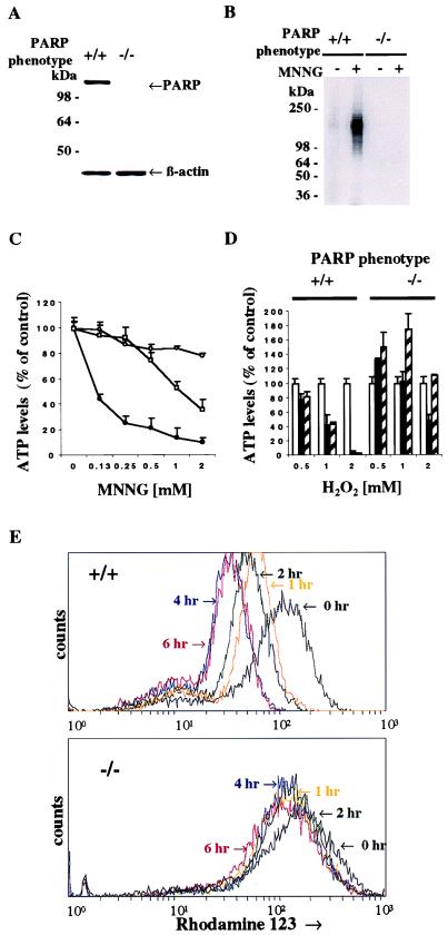
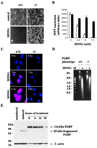
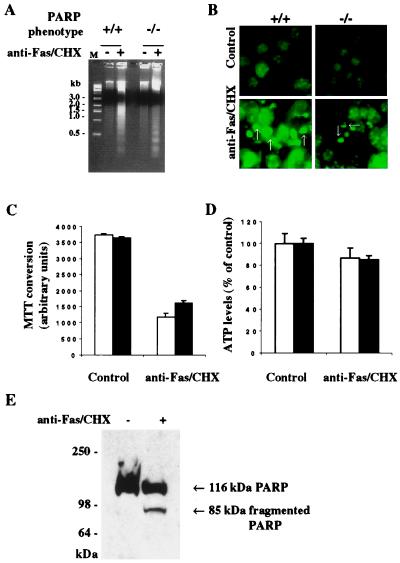
Apoptotic and necrotic cell death are well characterized and are influenced by intracellular ATP levels. Poly(ADP-ribose) polymerase (PARP), a nuclear enzyme activated by DNA strand breaks, physiologically participates in DNA repair. Overactivation of PARP after cellular insults can lead to cell death caused by depletion of the enzyme's substrate beta-nicotinamide adenine dinucleotide and of ATP. In this study, we have differentially elicited apoptosis or necrosis in mouse fibroblasts. Fibroblasts from PARP-deficient (PARP(-/-)) mice are protected from necrotic cell death and ATP depletion but not from apoptotic death. These findings, together with cell death patterns in PARP(-/-) animals receiving other types of insults, indicate that PARP activation is an active trigger of necrosis, whereas other mechanisms mediate apoptosis.


Figures

Figure 1
Figure 1
PARP activity regulates intracellular ATP levels. (A) Protein immunoblot analysis of PARP expression in PARP+/+ and PARP−/− MEFs. The PARP−/− lane confirms the absence of PARP protein in MEFs of PARP−/− mice. Total protein from PARP+/+ and PARP−/− MEFs was separated by SDS/12% PAGE. β-Actin (Sigma) was used as a loading control. (B) Activation of PARP catalytic activity in wild-type MEFs. Treatment with MNNG induced PARP catalytic activity in the PARP+/+ but not PARP−/− MEFs (arrow). Both PARP+/+ and PARP−/− MEFs were treated with 0.5 mM MNNG for 1 hr, and PARP catalytic activity was assayed as described (38). (C) Treatment with MNNG depleted intracellular ATP in wild-type but not PARP−/− MEFs. Solid circles, PARP+/+ MEFs; open circles, PARP−/− MEFs; open squares, PARP+/+ MEFs with 10 μM 3,4-dihydro-5-[4-(1-piperidinyl)butox]-1(2H)-isoquinolinone (DPQ). MNNG depleted intracellular ATP in a concentration-dependent manner. The depletion of intracellular ATP markedly diminished in PARP−/− and in PARP+/+ treated with DPQ. The data are presented as percentage of control content for each of two genotypes. The initial values of ATP level were 3.31 ± 0.36 nmol per 1 × 10⁶ cells for PARP+/+ and 3.50 ± 0.13 nmol per 1 × 10⁶ cells for PARP−/−. Intracellular ATP levels were determined by a luciferin/luciferase method. (D) H2O2 depleted ATP levels in wild-type but not PARP−/− MEFs. Open bar, 0 hr; solid bar, 1 hr; hatched bar, 2 hr. The H2O2-induced depletion of intracellular ATP was blocked in PARP−/− MEFs. Data are presented as percentage of control content for each of two untreated genotypes. (C–D) Both PARP+/+ and PARP−/− MEFs were treated with various doses of MNNG for 1 hr and H2O2 for 1 and 2 hr. PARP inhibitor, DPQ (10 μM), was pretreated for 3 hr before addition of MNNG. Data are means ± SD of triplicate determinations and representative of at least three experiments. (E) Loss of mitochondrial membrane potential in PARP+/+ MEFs treated with 5 mM MNNG. PARP+/+ MEFs lost the mitochondrial membrane potential in a time-dependent manner, whereas PARP−/− MEFs showed only slight loss of mitochondrial membrane potential after treatment of MNNG.
Figure 2
Figure 2
PARP overactivation induced by MNNG mediates necrosis. (A) Morphological profiles of MNNG-induced cell death in PARP+/+ MEFs. PARP+/+ MEFs treated with MNNG detached from culture plates, whereas PARP−/− MEFs treated with MNNG were protected from detachment and were morphologically similar to untreated MEFs. MEFs after treatment of 5 mM MNNG for 3 hr are shown in phase-contrast pictures. All photographs were taken at ×20 magnification. (B) Viability of MEFs treated with various concentrations of MNNG. Open bar, PARP+/+ MEFs; solid bar, PARP−/− MEFs. PARP+/+ MEFs lost cell viability, whereas PARP−/− MEFs maintained cell viability. Both PARP+/+ and PARP−/− MEFs were treated with various concentrations of MNNG for 6 hr. Cell viability was determined 6 hr later by formazan production from diphenyltetrazolium salt. Data are means ± SD of triplicate determinations and representative of at least three experiments. (C) Necrotic cell death induced by MNNG in PARP+/+ MEFs. Necrotic PARP+/+ MEFs had orange-red fluorescence, because propidium iodide gained access to the nucleus. Viable PARP−/− MEFs had blue fluorescence, because only membrane-permeable Hoechst 33324 gained access to the nucleus, as in untreated MEFs. Both PARP+/+ and PARP−/− MEFs were loaded with propidium iodide (10 μg/ml) and Hoechst 33342 (10 μg/ml) after 0, 3, and 6 hr after treatment with 5 mM MNNG. (D) Lack of PARP cleavage in PARP+/+ MEFs treated with 5 mM MNNG. PARP+/+ MEFs were treated with 5 mM MNNG for 0, 6, 12, and 18 hr. Total protein from PARP+/+ MEFs was separated by SDS/4–12% PAGE. β-actin was used as a loading control. M, 1-kilobase DNA marker (39). (E) MNNG-induced necrotic DNA damage. Treatment of MEFs with 5 mM MNNG for 6 hr resulted in a DNA smear.
Figure 3
Figure 3
Fas-induced apoptosis without ATP depletion. (A) Fas-induced internucleosomal DNA fragmentation. DNA laddering was essentially the same in PARP+/+ and PARP−/− MEFs treated with anti-Fas and CHX for 24 hr (39). M, 1-kilobase DNA marker. (B) Fas-induced nuclear fragmentation. Nuclear fragmentation (arrows) occurred in both PARP+/+ and PARP−/− MEFs treated with anti-Fas and CHX for 24 hr. After the treatments with anti-Fas and CHX, MEFs were stained with Hoechst 33342. Hoechst 33342-stained apoptotic nuclei had fragmentation with pseudocolored green fluorescence. All photographs were taken at ×20 magnification. (C) Fas-induced cell death. Open bar, PARP+/+ MEFs; solid bar, PARP−/− MEFs. Both PARP+/+ and PARP−/− MEFs were susceptible to apoptotic cell death induced by anti-Fas and CHX. Cell viability was determined 24 hr later by formazan production from diphenyltetrazolium salt. Data are means ± SD of triplicate determinations and representative of at least three experiments. (D) Effects of Fas on intracellular ATP levels. Intracellular ATP levels were maintained in both PARP+/+ and PARP−/− MEFs treated with anti-Fas and CHX. Open bar, PARP+/+ MEFs; solid bar, PARP−/− MEFs. The data are presented as percentage of control content for each of two genotypes. The intracellular ATP levels were determined by a luciferin/luciferase method. (E) Fas-induced cleavage of PARP in PARP+/+ MEFs. Total proteins from treated and untreated PARP+/+ MEFs were separated by SDS/4–12% PAGE. (A–E) Both PARP+/+ and PARP−/− MEFs were treated with anti-Fas (100 ng/ml) and CHX (10 μg/ml) for 24 hr.

References

    1. Lautier D, Lagueux J, Thibodeau J, Menard L, Poirier G G. Mol Cell Biochem. 1993;122:171–193. - PubMed
    1. de Murcia G, Schreiber V, Molinete M, Saulier B, Poch O, Masson M, Niedergang C, Menissier de Murcia J. Mol Cell Biochem. 1994;138:15–24. - PubMed
    1. Satoh M S, Lindahl T. Nature (London) 1992;356:356–358. - PubMed
    1. Berger N A. Radiat Res. 1985;101:4–15. - PubMed
    1. Carson D A, Seto S, Wasson D B, Carrera C J. Exp Cell Res. 1986;164:273–281. - PubMed

Publication types

LinkOut - more resources