. 1999 Nov 23;96(24):14037-42.
doi: 10.1073/pnas.96.24.14037.

Differentially expressed protein Pdcd4 inhibits tumor promoter-induced neoplastic transformation

Affiliations

Differentially expressed protein Pdcd4 inhibits tumor promoter-induced neoplastic transformation

J L Cmarik et al. Proc Natl Acad Sci U S A. .

Abstract

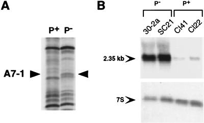
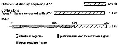
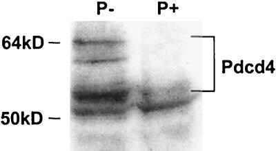
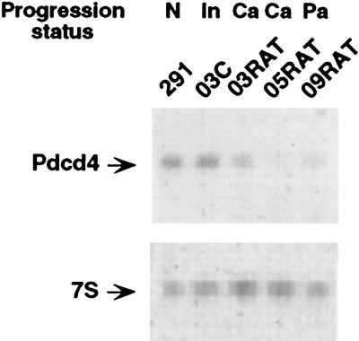
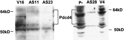
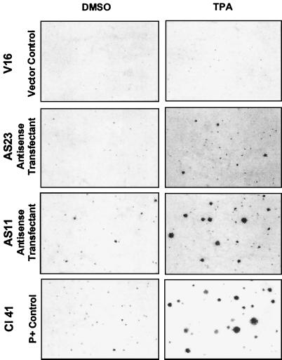
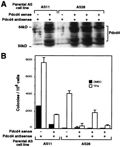
An mRNA differential display comparison of mouse JB6 promotion-sensitive (P+) and -resistant (P-) cells identified a novel gene product that inhibits neoplastic transformation. The JB6 P+ and P- cells are genetic variants that differ in their transformation response to tumor promoters; P+ cells form anchorage-independent colonies that are tumorigenic, and P- cells do not. A differentially displayed fragment, A7-1, was preferentially expressed in P- cells at levels >/=10-fold those in P+ cells, making its mRNA a candidate inhibitor of neoplastic transformation. An A7-1 cDNA was isolated that was identical to murine Pdcd4 gene cDNAs, also known as MA-3 or TIS, and analogous to human H731 and 197/15a. Until now, the function of the Pdcd4 protein has been unknown. Paralleling the mRNA levels, Pdcd4 protein levels were greater in P- than in P+ cells. Pdcd4 mRNA was also expressed at greater levels in the less progressed keratinocytes of another mouse skin neoplastic progression series. To test the hypothesis that Pdcd4 inhibits tumor promoter-induced transformation, stable cell lines expressing antisense Pdcd4 were generated from parental P- cells. The reduction of Pdcd4 proteins in antisense lines was accompanied by acquisition of a transformation-sensitive (P+) phenotype. The antisense-transfected cells were reverted to their initial P- phenotype by overexpression of a Pdcd4 sense fragment. These observations demonstrate that the Pdcd4 protein inhibits neoplastic transformation.


Figures

Figure 1
Figure 1
Differential display fragment A7-1 is preferentially expressed in JB6 P− cells. (A) Differential display with T12MA and AP-7 primers was carried out on total RNA from P+ and P− cells and separated on a 6% denaturing polyacrylamide gel as described in Materials and Methods. Arrowheads indicate the position of a band, A7-1, reproducibly visible only in the P− lane. One set of duplicate samples is shown. (B) Northern analysis of total RNA (20 μg) prepared from untreated monolayer cultures of two independently derived P− cell lines (30–2a and SC21) and two independently derived P+ cell lines (Cl41 and Cl22) was carried out by using a 32P-labeled A7-1 probe (upper panel). The arrow indicates the approximate size of the A7-1 band. The blot was stripped and reprobed with a 32P-labeled 7S RNA probe to assess equivalent loading (lower panel).
Figure 2
Figure 2
Alignment of A7-1 differential display fragment and cDNAs. Identical regions of the A7-1 fragment, the P− cDNA clone, and the sequence of MA-3 reported by Shibahara et al. (22) are indicated by the hatched regions. The A7-1 P− clone starts at nucleotide 1023 of MA-3. The ORF of the full-length clone is indicated by the shaded region and extends from nucleotides 71 to 1478. The black box near the 5′-end of the full-length clone indicates the putative nuclear localization signal.
Figure 3
Figure 3
Preferential expression of Pdcd4 proteins in P− cells. Lysates were prepared from 30–2a P− cells and Cl22 P+ cells. Proteins (12 μg) were separated on 10% Bis-Tris NuPage gels, transferred to nitrocellulose, and subjected to immunoblotting with anti-H731 polyclonal antibody with visualization by chemiluminescent detection. The migration of molecular weight standards is indicated.
Figure 4
Figure 4
Preferential expression of Pdcd4 mRNA in preneoplastic lines of mouse keratinocyte progression series. Northern analysis of total RNA (10 μg) prepared from normal (N) cell line 291, initiated (In) cell line 03C, carcinoma (Ca) cell line 03RAT, Ca cell line 05RAT, and papilloma (Pa) cell line 09RAT was carried out by using a 32P-labeled Pdcd4 probe (upper panel). The blot was stripped and reprobed with a 32P-labeled 7S RNA probe to assess equivalent loading (lower panel).
Figure 5
Figure 5
Antisense expression reduces Pdcd4 protein levels. Lysates were prepared from independent transfectant cell line clones (vector controls V16 and V4 and Pdcd4 antisense transfectants AS11, AS23, and AS28) and 30–2a P− cells, and immunoblotting was carried out as described for Fig. 3. The migration of molecular weight standards is indicated.
Figure 6
Figure 6
Gain of transformation response in P− cells expressing antisense Pdcd4. Cell lines were exposed to the solvent control 0.1% DMSO or to 10 ng/ml (16 nM) of TPA for 14 days in soft agar. Dishes were stained with 2-(4-iodophenyl)-3-(4-nitrophenyl)-5-phenyltetrazolium chloride and photographed at 25× magnification. Examples representative of several experiments are shown.
Figure 7
Figure 7
Pdcd4 sense fragment abrogates antisense effect. Pooled transfectant cell lines with or without the Pdcd4 sense fragment were established from the parental antisense lines AS11 and AS28. (A) Immunoblotting with anti-H731 was carried out as described for Fig. 3. The migration of molecular weight standards is indicated. Coomassie blue staining indicated that the protein amount in each lane varied by <20%. (B) An anchorage-independent transformation assay was carried out as described for Fig. 6, and the colony number was determined with a computerized image analyzer. Error bars indicate the range of values of duplicate dishes.

References

    1. Liang P, Pardee A B. Science. 1992;257:967–971. - PubMed
    1. Herschman H R. In: Advances in Regulation of Cell Growth, Volume 2; Cell Activation: Genetic Approaches. Mond J J, Cambier J C, Weiss A, editors. New York: Raven; 1991. pp. 83–117.
    1. Cmarik J L, Colburn N H. In: Food Factors for Cancer Prevention. Ohigashi H, Osawa T, Terao J, Watanabe S, Yoshikawa T, editors. Tokyo: Springer-Verlag; 1997. pp. 67–76.
    1. Colburn N H, Former B F, Nelson K A, Yuspa S H. Nature (London) 1979;281:589–591. - PubMed
    1. Colburn N H, Wendel E, Abruzzo G. Proc Natl Acad Sci USA. 1981;78:6912–6916. - PMC - PubMed

Publication types

LinkOut - more resources