. 1999 Dec;2(12):1131-6.
doi: 10.1038/16056.

Dual streams of auditory afferents target multiple domains in the primate prefrontal cortex

Affiliations

Dual streams of auditory afferents target multiple domains in the primate prefrontal cortex

L M Romanski et al. Nat Neurosci. 1999 Dec.

Abstract

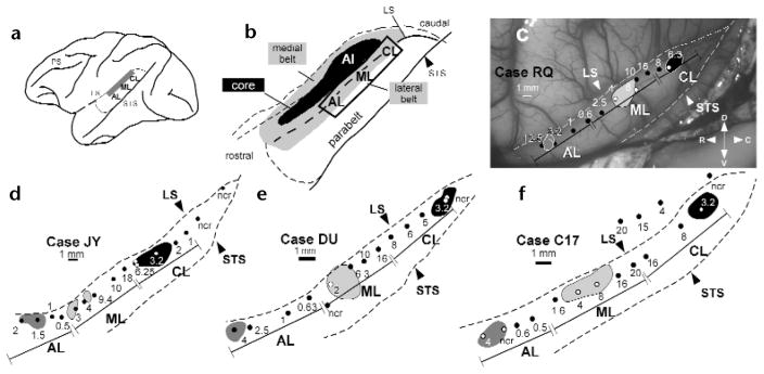
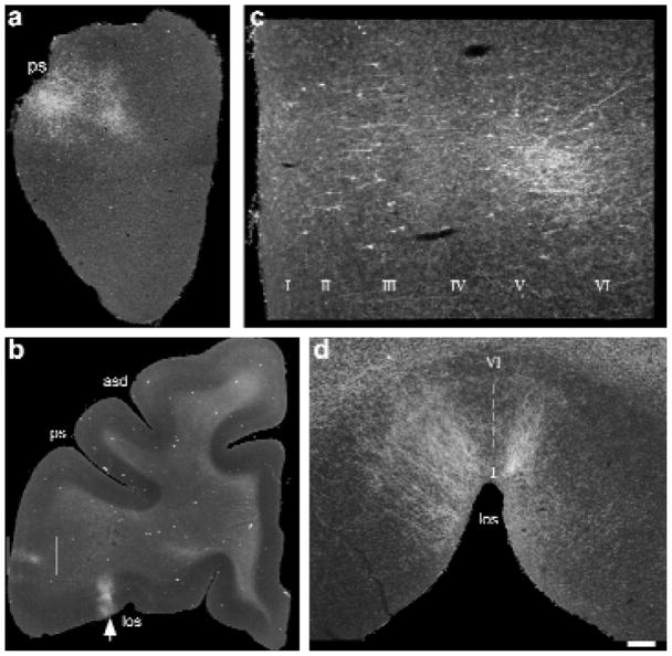
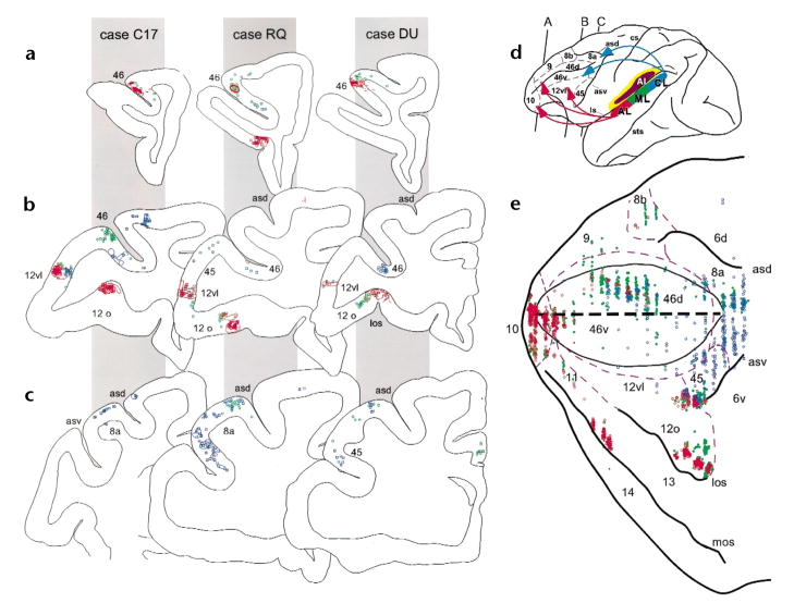
'What' and 'where' visual streams define ventrolateral object and dorsolateral spatial processing domains in the prefrontal cortex of nonhuman primates. We looked for similar streams for auditory-prefrontal connections in rhesus macaques by combining microelectrode recording with anatomical tract-tracing. Injection of multiple tracers into physiologically mapped regions AL, ML and CL of the auditory belt cortex revealed that anterior belt cortex was reciprocally connected with the frontal pole (area 10), rostral principal sulcus (area 46) and ventral prefrontal regions (areas 12 and 45), whereas the caudal belt was mainly connected with the caudal principal sulcus (area 46) and frontal eye fields (area 8a). Thus separate auditory streams originate in caudal and rostral auditory cortex and target spatial and non-spatial domains of the frontal lobe, respectively.


Figures

Fig. 1
Fig. 1
Tonotopic maps of the lateral belt region recorded in four monkeys. (a) Lateral belt region (shaded in gray) overlies the superior temporal plane and dorsal aspect of the superior temporal gyrus. (b) The same region, enlarged to show the locations of the medial and lateral belt (shaded in gray), primary auditory cortex (AI, black) and the parabelt cortex on the superior temporal gyrus. The portion of the lateral belt recorded from is outlined. (c–f) Physiological maps of the lateral belt recordings from each of the four monkeys in a schematic of the superior temporal region. (c) Photomicrograph taken during the electrophysiological recordings of the lateral belt in case RQ. (d–f) Mappings from the other three cases. The best center frequency for each electrode penetration (black or white dots) is labeled in kHz. Injections of different anterograde and retrograde tracers (shaded regions) for each case are shown with respect to these recordings (for details of tracers in each case see Methods). The boundaries of AL, ML and CL are delineated by a bounded line and are derived from the frequency reversal points. Abbreviations: ncr, no clear response for this electrode penetration, LS, lateral sulcus; PS, principal sulcus; STS, superior temporal sulcus; D, dorsal; V, ventral; R, rostral; C, caudal.
Fig. 2
Fig. 2
Photomicrographs of anterograde and retrograde labeling in the rostral and ventral regions of the prefrontal cortex after injections of Fluoro-ruby (a, d; case DU) or Fluoro-Emerald (b, c; case RQ) into area AL. (a) Low-power photomicrograph of anterogradely labeled fibers in a coronal section through the frontal pole region, just anterior to the beginning of the principal sulcus. (b) Low-power photomicrograph from a coronal section with labeling in the lateral inferior convexity (boxed region) and the lateral orbital cortex (arrow). (c) The boxed region at higher power. Cells in layers III and V were retrogradely labeled, whereas anterogradely labeled fibers were seen in layers II, III, V and VI. (d) Photomicrograph of the same lateral orbital region (b; arrow) from a different case (DU), detailing the lateral orbital projections at higher magnification in a coronal section in which they were densest. Labeled axons made columnar terminations spanning all layers (I–VI) at the juncture of the lateral orbital sulcus. Scale bar (a) 600 μm; (b) 2250 μm; (c)250 μm; (d) 425 μm. Abbreviations: asd, dorsal arcuate sulcus; los, lateral orbital sulcus; ps, principal sulcus.
Fig. 3
Fig. 3
Anterograde and retrograde labeling in the prefrontal cortex. In three monkeys (cases C17, RQ and DU indicated by gray panels) coronal sections from anterior (a), middle (b) and posterior (c) levels of the prefrontal cortex are shown with color coded retrograde cells (dots and squares) and anterograde fibers (outlined shapes). The cytoarchitectonic region is shown next to the labeling in each coronal section. Across all cases, projections from injections into AL (red) were found in the frontal pole (not shown), the rostral principal sulcus (level a) and in the lateral inferior convexity and lateral orbital cortex (level b). In contrast, projections from CL (blue) can be seen in more caudal principal sulcus regions in (b) as well as in the periarcuate cortex shown in (c)and also in the lateral inferior convexity in (b). Projections from area ML, shown in green, overlapped with projections from AL and CL. (d) Lateral brain schematic showing color coding of the lateral belt injections and the cytoarchitectonic organization of the prefrontal cortex as well as the rostrocaudal levels of the coronal sections from (a–c). Projections are summarized with arrows. (e) Flattened map of dorsolateral and ventral prefrontal cortex showing the projections from case RQ shows that AL projections (red) target 10, rostral 46 and ventral prefrontal regions (areas 12vl, l2o and 45) whereas CL (blue) targets the caudal principal sulcus and periarcuate regions (caudal 46 and 8a) and the lateral inferior convexity (areas 12vl and 45). The dashed purple lines indicate described cytoarchitectonic borders and edges of major sulci are shown in black. The center black dashed line represents the principal sulcus.

Comment in

References

    1. Ungerleider LG, Mishkin M. In: Analysis of Visual Behavior. Ingle DJ, Goodale MA, Mansfield RJW, editors. MIT Press; Cambridge, Massachusetts: 1982. pp. 549–586.
    1. Wilson FA, O’Scalaidhe SP, Goldman-Rakic PS. Dissociation of object and spatial processing domains in primate prefrontal cortex. Science. 1993;260:1955–1958. - PubMed
    1. Webster MJ, Bachevalier J, Ungerleider LG. Connections of inferior temporal areas TEO and TE with parietal and frontal cortex in macaque monkeys. Cereb Cortex. 1994;4:470–483. - PubMed
    1. Buckner RL, Raichle ME, Petersen SE. Dissociation of human prefrontal cortical areas across different speech production tasks and gender groups. J Neurophysiol. 1995;74:2163–2173. - PubMed
    1. Stromswold K, Caplan D, Alpert N, Rauch S. Localization of syntactic comprehension by positron emission tomography. Brain Lang. 1996;52:452–473. - PubMed

Substances