. 1999 Dec;42(6):1048-60.
doi: 10.1002/(sici)1522-2594(199912)42:6<1048::aid-mrm9>3.0.co;2-m.

Cardiac motion tracking using CINE harmonic phase (HARP) magnetic resonance imaging

Affiliations

Cardiac motion tracking using CINE harmonic phase (HARP) magnetic resonance imaging

N F Osman et al. Magn Reson Med. 1999 Dec.

Abstract

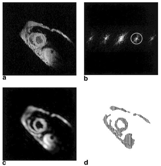
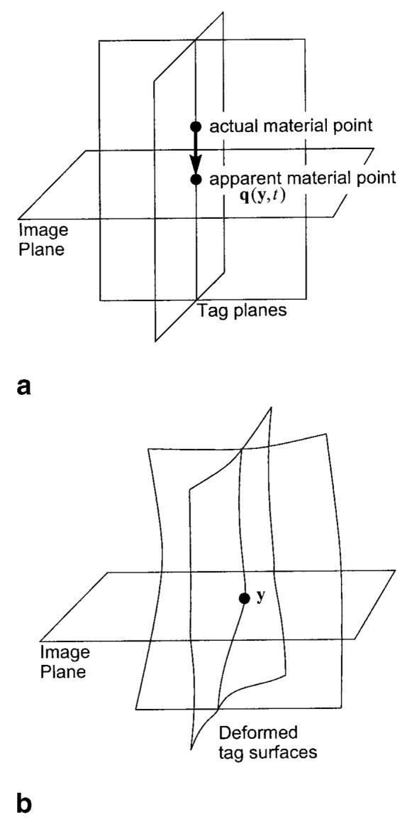
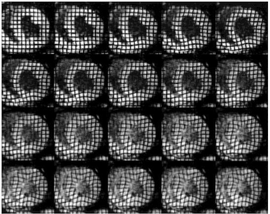
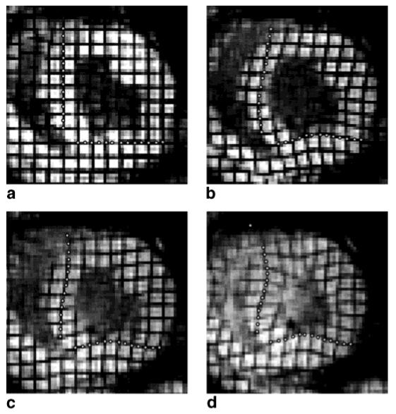
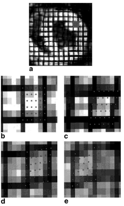
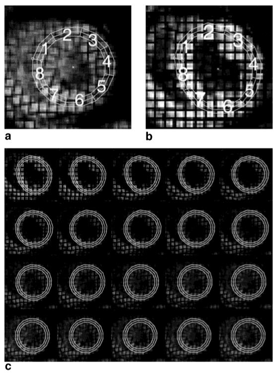
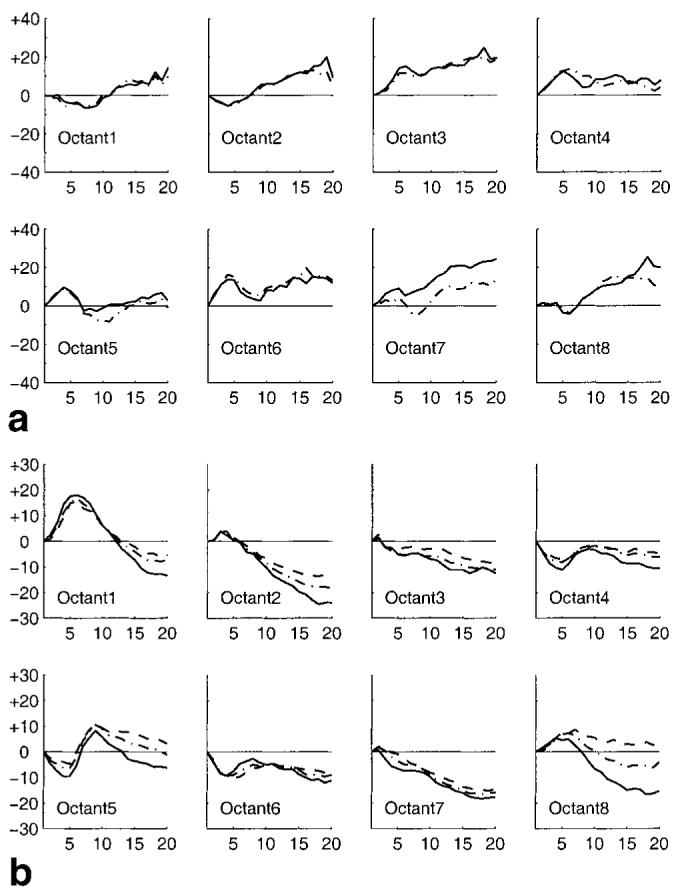
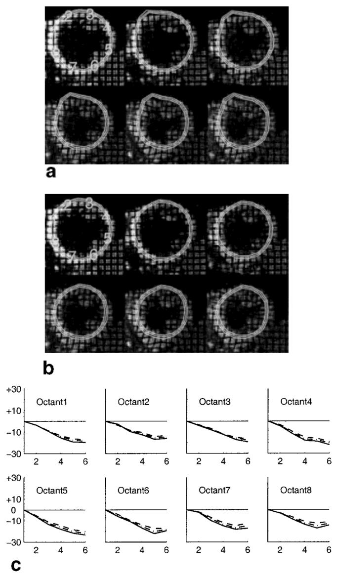
This article introduces a new image processing technique for rapid analysis of tagged cardiac magnetic resonance image sequences. The method uses isolated spectral peaks in SPAMM-tagged magnetic resonance images, which contain information about cardiac motion. The inverse Fourier transform of a spectral peak is a complex image whose calculated angle is called a harmonic phase (HARP) image. It is shown how two HARP image sequences can be used to automatically and accurately track material points through time. A rapid, semiautomated procedure to calculate circumferential and radial Lagrangian strain from tracked points is described. This new computational approach permits rapid analysis and visualization of myocardial strain within 5-10 min after the scan is complete. Its performance is demonstrated on MR image sequences reflecting both normal and abnormal cardiac motion. Results from the new method are shown to compare very well with a previously validated tracking algorithm. Magn Reson Med 42:1048-1060, 1999.


Figures

FIG. 1
FIG. 1
a: An MR image with vertical SPAMM tags. b: Shows the magnitude of its Fourier transform. By extracting the spectral peak inside the circle in b, a complex image is produced with a magnitude (c) and a phase (d).
FIG. 2
FIG. 2
a: Tag planes at end-diastole. b: distortion of the tag planes due to motion.
FIG. 3
FIG. 3
Concentric circles within the LV wall and eight octants.
FIG. 4
FIG. 4
A sequence of tagged MR images of a paced canine heart composed by multiplying the vertical and horizontal tag images to show a grid. The images are 20 time frames depicting the motion of a paced canine heart from end-diastole (top left) to end-systole (bottom right).
FIG. 5
FIG. 5
(a) Manually selected points at time frame 1 are tracked through time and displayed at time frames (b) 5, (c) 10, and (d) 20.
FIG. 6
FIG. 6
(a) The heart at end-diastole showing the position of the densely picked points; an enlargement of the tracked material points at time frames 1 (b), 5 (c), 10 (d), and 20 (e).
FIG. 7
FIG. 7
a: Manually defined circles at end-systole. b: The deformed shape of these circles after tracking backwards to end-diastole. c: The entire sequence of tracked circles.
FIG. 8
FIG. 8
a: The time evolution of epicardial (dot-dashed) and endocardial (solid) radial strain in each octant. b: The time evolution of epicardial (dashed), midwall (dot-dashed), and endocardial (solid) circumferential strain in each octant.
FIG. 9
FIG. 9
Results from a normal human heart undergoing dobutamine induced stress. a: Short-axis images and tracked circles from first (top-left) to last (bottom-right) without HARP refinement. b: The result after application of HARP refinement. c: The temporal evolution of circumferential strain calculated using the refined points.
FIG. 10
FIG. 10
HARP accuracy in comparison to FindTags using normal human data. a: A tagged image with tag points from FindTags (black dots) overlaying HARP π isocontours (white curves). b: Average distance between the FindTags tag points and the HARP isocontour.
FIG. 11
FIG. 11
Root mean square difference in tag crossing estimation between Findtags and HARP.

References

    1. Zerhouni EA, Parish DM, Rogers WJ, Yang A, Shapiro EP. Human heart: tagging with MR imaging—a method for noninvasive assessment of myocardial motion. Radiology. 1988;169(1):59–63. - PubMed
    1. Axel L, Dougherty L. MR imaging of motion with spatial modulation of magnetization. Radiology. 1989;171:841–845. - PubMed
    1. McVeigh ER, Atalar E. Cardiac tagging with breath-hold cine MRI. Magn Reson Med. 1992;28:318–327. - PMC - PubMed
    1. Fischer SE, McKinnon GC, Maier SE, Boesiger P. Improved myocardial tagging contrast. Magn Reson Med. 1993;30:191–200. - PubMed
    1. Atalar E, McVeigh ER. Minimization of dead-periods in MRI pulse sequences for imaging oblique planes. Magn Reson Med. 1994;32(6):773–777. - PMC - PubMed

Publication types

LinkOut - more resources