. 1999 Dec;181(23):7285-90.
doi: 10.1128/JB.181.23.7285-7290.1999.

A periplasmic location is essential for the role of the ApbE lipoprotein in thiamine synthesis in Salmonella typhimurium

Affiliations

A periplasmic location is essential for the role of the ApbE lipoprotein in thiamine synthesis in Salmonella typhimurium

B J Beck et al. J Bacteriol. 1999 Dec.

Abstract

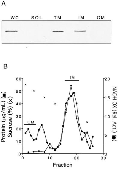
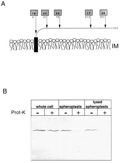
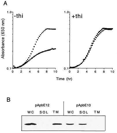
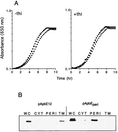
ApbE is a lipoprotein in Salmonella typhimurium, and mutants unable to make this protein have a reduced ability to make thiamine (vitamin B(1)) and require it as a supplement for optimal growth in minimal glucose medium. Polyclonal antibodies specific to ApbE were used to determine that wild-type ApbE is located exclusively in the inner membrane. The periplasmic, monotopic topology of ApbE was determined by using computer-based hydrophobicity plots, LacZ and PhoA gene fusions, and proteinase protection experiments. This extracellular location of ApbE is required for its function, since a cytoplasmic form (ApbE(cyto)) did not allow an apbE mutant to grow in the absence of thiamine. A periplasmic form of ApbE (ApbE(peri)) lacking the lipoprotein modification allowed an apbE mutant to grow in the absence of thiamine, indicating that soluble ApbE could function in thiamine synthesis and that lipoation and membrane association were not required. Alteration of the amino acid implicated in membrane sorting for other lipoproteins did not result in a relocalization of ApbE to the outer membrane, suggesting that additional sorting determinants exist for ApbE.


Figures

FIG. 1
FIG. 1
Chromosomally encoded ApbE from S. typhimurium LT2 is located in the inner membrane. (A) Immunoblot of whole-cell (WC), soluble cytoplasmic and periplasmic (SOL), total membrane (TM), inner membrane (IM), and outer membrane (OM) fractions. Cellular fractions were prepared as described in Materials and Methods. Western analysis with polyclonal antibodies against ApbE (1:5,000 dilution) was performed (14). (B) Representative membrane separation profile from S. typhimurium LT2. Inner and outer membranes were fractionated by sucrose density equilibration. The protein concentration, NADH oxidase activity, and sucrose concentration values are shown. The fractions pooled and used in Fig. 1A are indicated.
FIG. 2
FIG. 2
ApbE is periplasmically located. (A) The predicted monotopic structure of ApbE anchored in the outer leaflet of the inner membrane lipid bilayer is schematically shown. Arrows indicate the location of active alkaline phosphatase (PhoA) fusions to ApbE encoded by pApbE1, and the number below the shaded box indicates the amino acid of ApbE that immediately proceeds the fusion junction. The numbers in the shaded boxes represent the relative PhoA activity (in units per minute per optical density at 650 nm) of each fusion compared to a control containing an out-of-frame PhoA fusion. (B) Immunoblot showing the proteinase K accessibility of chromosomally encoded ApbE in whole cells, spheroplasts, and lysed spheroplasts. Samples were treated with (+) or without (−) proteinase K (100 μg/ml). The proteins in each sample (equivalent to 0.05 A650) were separated by SDS-PAGE and immunoblotted with a 1:5,000 dilution of ApbE antisera.
FIG. 3
FIG. 3
ApbE requires a signal peptide for membrane association and function in thiamine synthesis. (A) The growth of strain DM271 (apbE) carrying pApbE12, encoding wild-type ApbE (●), or pApbE10, encoding ApbE without the lipoprotein signal peptide (■), is shown. Cells were grown in minimal medium with glucose as the sole carbon and energy source and supplemented with 100 nM thiamine where indicated. (B) Immunoblot showing the cellular location of ApbE produced by pApbE10 and pApbE12. Cellular fractions were isolated as described in Materials and Methods. Abbreviations: WC, whole-cell fraction; SOL, soluble cytoplasmic and periplasmic proteins; TM, total membrane proteins.
FIG. 4
FIG. 4
A periplasmic form of ApbE complements the thiamine requirement of an apbE mutant. (A) Growth of strain DM271 (apbE) carrying pApbEperi (●) or pApbE12 that produces wild-type ApbE (■) in minimal glucose medium supplemented with 10 nM thiamine where indicated. (B) Immunoblot showing the cellular location of ApbE and ApbEperi expressed from pApbE12 and pApbEperi, respectively. Cellular fractions were isolated as described in Materials and Methods. Abbreviations: WC, whole-cell proteins; CYT, cytoplasmic proteins; PERI, periplasmic proteins; TM, total membrane proteins.

References

    1. Barik S. Site-directed mutagenesis by double polymerase chain reaction. Mol Biotechnol. 1995;3:1–7. - PubMed
    1. Bartolomé B, Jubete Y, Martinez E, de la Cruz F. Construction and properties of a family of pACYC184-derived cloning vectors compatible with pBR322 and its derivatives. Gene. 1991;102:75–78. - PubMed
    1. Beck B J, Connolly L E, de las Penas A, Downs D M. Evidence that rseC, a gene in the rpoE cluster, has a role in thiamine synthesis in Salmonella typhimurium. J Bacteriol. 1997;179:6504–6508. - PMC - PubMed
    1. Beck B J, Downs D M. The apbE gene encodes a lipoprotein involved in thiamine synthesis in Salmonella typhimurium. J Bacteriol. 1998;180:885–891. - PMC - PubMed
    1. Begley T P, Downs D M, Ealick S, McLafferty F, van Loon D, Taylor S, Chiu H, Kinsland C, Reddick J, Xi J, Campobasso N. Thiamin synthesis in prokaryotes. Arch Microbiol. 1999;171:293–300. - PubMed

Publication types

MeSH terms

LinkOut - more resources