. 1999 Dec;181(23):7308-13.
doi: 10.1128/JB.181.23.7308-7313.1999.

Molecular evidence for a new bacteriophage of Borrelia burgdorferi

Affiliations

Molecular evidence for a new bacteriophage of Borrelia burgdorferi

C H Eggers et al. J Bacteriol. 1999 Dec.

Abstract

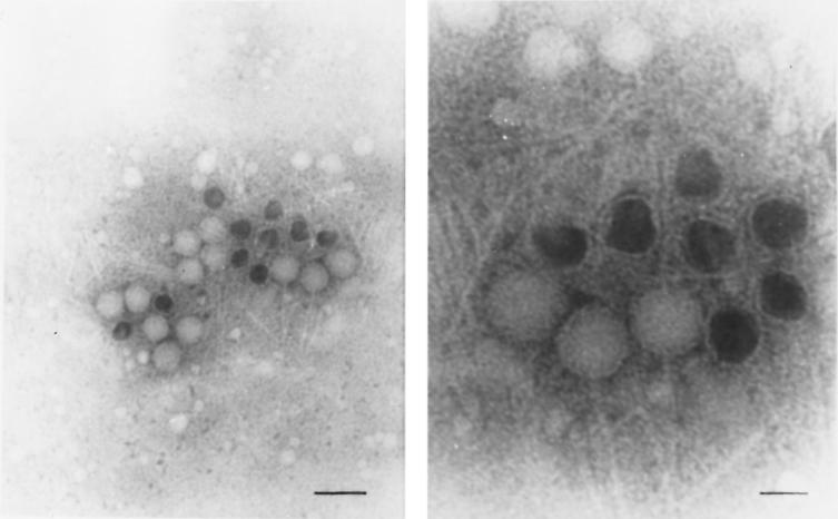
We have recovered a DNase-protected, chloroform-resistant molecule of DNA from the cell-free supernatant of a Borrelia burgdorferi culture. The DNA is a 32-kb double-stranded linear molecule that is derived from the 32-kb circular plasmids (cp32s) of the B. burgdorferi genome. Electron microscopy of samples from which the 32-kb DNA molecule was purified revealed bacteriophage particles. The bacteriophage has a polyhedral head with a diameter of 55 nm and appears to have a simple 100-nm-long tail. The phage is produced constitutively at low levels from growing cultures of some B. burgdorferi strains and is inducible to higher levels with 10 microg of 1-methyl-3-nitroso-nitroguanidine (MNNG) ml(-1). In addition, the prophage can be induced with MNNG from some Borrelia isolates that do not naturally produce phage. We have isolated and partially characterized the phage associated with B. burgdorferi CA-11.2A. To our knowledge, this is the first molecular characterization of a bacteriophage of B. burgdorferi.


Figures

FIG. 1
FIG. 1
DNase protection of extracellular bacteriophage DNA. After PEG precipitation, phage samples were extracted twice with chloroform. Samples were subjected to digestion with DNase I prior to DNA extraction. After DNA isolation, the samples either were loaded onto a 0.8% agarose gel (lane 1) or were subjected to another digestion with DNase I and then loaded directly onto the agarose gel (lane 2). The DNA was resolved with field inversion gel electrophoresis. The gel was stained with EtBr. Molecular sizes are in kilobase pairs.
FIG. 2
FIG. 2
Denaturation of phage DNA. DNA denatured with 0.2 M NaOH (+) and an untreated control (−) were resolved on a 0.5% agarose gel. The denatured phage DNA did not “snap back” and regenerate a double-stranded DNA molecule, as did the denatured covalently closed linear plasmid lp17. The arrow indicates single-stranded DNA products generated by the denaturation of non-covalently closed double-stranded DNA. The gel was stained with EtBr. Molecular sizes are in kilobase pairs.
FIG. 3
FIG. 3
Genomic location of prophage DNA. Total cellular DNA from B. burgdorferi CA-11.2A was resolved by two-dimensional gel electrophoresis (left). The large circular plasmid (white arrow) was retarded in its migration in the second dimension, and the linear plasmids migrated on the diagonal. A Southern blot of the gel was probed with total phage DNA that was extracted and radiolabeled (right). The phage DNA hybridized to the circular 32-kb plasmid (black arrow). Additionally, the phage DNA hybridized to the nicked (upper band) and linearized (middle band) forms of cp32 that were generated during DNA extraction. Molecular sizes are in kilobase pairs.
FIG. 4
FIG. 4
Induction of prophage from different Borrelia strains. Phage DNA was extracted from 10 ml of cell-free supernatants from B. burgdorferi B31 (lanes 1 to 3), B. burgdorferi CA-11.2A (lanes 4 to 6), and B. bissettii DN127 (lanes 7 to 9) cultures. The DNA was collected from log-phase starter cultures (lanes 1, 4, and 7), untreated controls (lanes 2, 5, and 8), and cultures treated with 10 μg of MNNG ml−1 (lanes 3, 6, and 9) and was electrophoresed on a 0.5% agarose gel. The gel was blotted and probed with cp32-specific probe 4 to enhance detection of phage DNA.
FIG. 5
FIG. 5
Microscopy of B. burgdorferi phage particles. Samples were collected from PEG-precipitated cell-free supernatants of an induced culture of B. burgdorferi CA-11.2A and viewed by TEM. (Left) Tailless heads, headless tails, and intact phage particles are visible, including both full and empty heads. Phosphotungstic acid stain; magnification, ×75,000 (bar = 150 nm). (Right) Close-up of the intact phage particles. Phosphotungstic acid stain; magnification, ×250,000 (bar = 40 nm).

References

    1. Ackerman H-W. Tailed bacteriophages: the order caudovirales. Adv Virus Res. 1998;51:135–201. - PMC - PubMed
    1. Amla D V. Mutagenesis of free and intracellular cyanophage AS-1 by ultraviolet, N-methanyl-N′-nitro-N-nitrosoguanidine and acriflavin. Mutat Res. 1979;59:147–155. - PubMed
    1. Balganesh M, Setlow J K. Prophage induction in Haemophilus influenzae and its relationship to mutation by chemical and physical agents. Mutat Res. 1984;125:15–22. - PubMed
    1. Barbour A G, Garon C F. Linear plasmids of the bacterium Borrelia burgdorferi have covalently closed ends. Science. 1987;237:409–411. - PubMed
    1. Barbour A G, Hayes S F. Biology of Borrelia species. Microbiol Rev. 1986;50:381–400. - PMC - PubMed

Publication types

MeSH terms

LinkOut - more resources