. 1999 Dec 1;19(23):10438-50.
doi: 10.1523/JNEUROSCI.19-23-10438.1999.

The contribution of facilitation of monosynaptic PSPs to dishabituation and sensitization of the Aplysia siphon withdrawal reflex

Affiliations

The contribution of facilitation of monosynaptic PSPs to dishabituation and sensitization of the Aplysia siphon withdrawal reflex

I Antonov et al. J Neurosci. .

Abstract

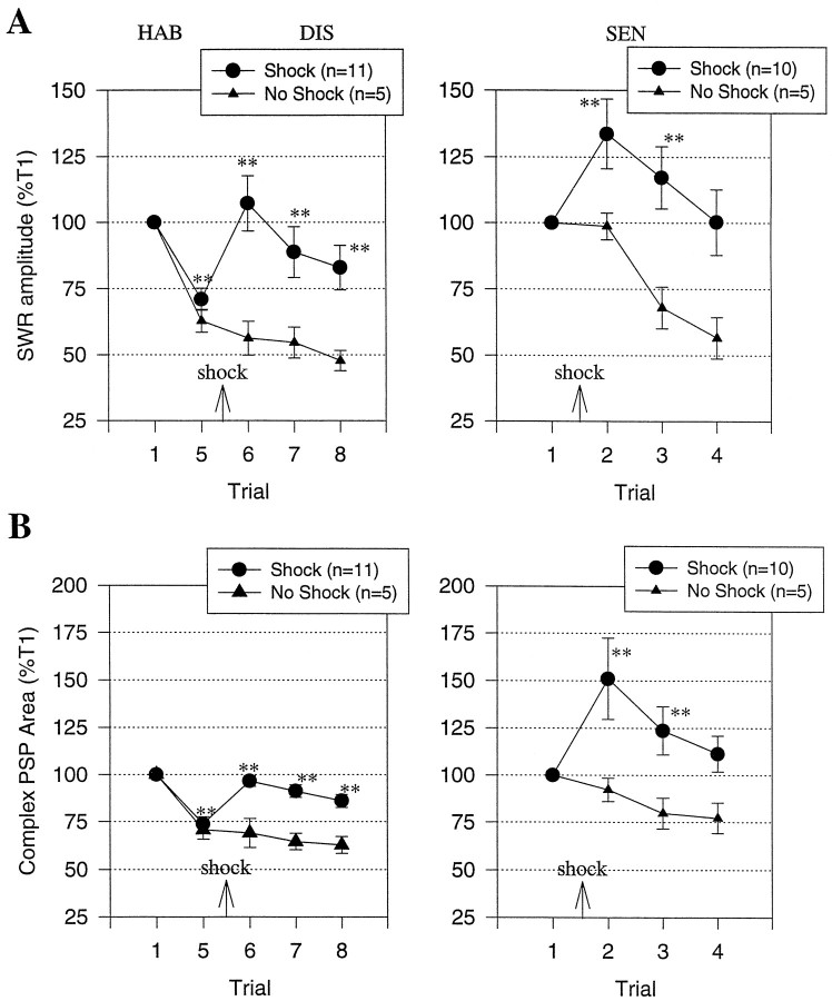
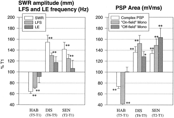
To examine the relationship between synaptic plasticity and learning and memory as directly as possible, we have developed a new simplified preparation for studying the siphon-withdrawal reflex of Aplysia in which it is relatively easy to record synaptic connections between individual identified neurons during simple forms of learning. We estimated that monosynaptic EPSPs from LE siphon sensory neurons to LFS siphon motor neurons mediate approximately one-third of the reflex response measured in this preparation, which corresponds to siphon flaring in the intact animal. To investigate cellular mechanisms contributing to dishabituation and sensitization, we recorded evoked firing of LFS neurons, the siphon withdrawal produced by stimulation of an LFS neuron, the complex PSP in an LFS neuron, and the monosynaptic PSP from an "on-field" or "off-field" LE neuron to an LFS neuron during behavioral training. Unlike the simplified gill-withdrawal preparation (Cohen et al., 1997; Frost et al., 1997), in the siphon-withdrawal preparation we found no qualitative differences between the major cellular mechanisms contributing to dishabituation and sensitization, suggesting that dissociations that have been observed previously may be attributable to transient inhibition that does not occur for this component of the reflex. Furthermore, in the siphon-withdrawal preparation, all of the various cellular measures, including monosynaptic PSPs from either on-field or off-field LE neurons, changed approximately in parallel with changes in the behavior. These results provide the most direct evidence so far available that both dishabituation and sensitization involve multiple mechanisms, including heterosynaptic facilitation of sensory neuron-motor neuron PSPs.


Figures

Fig. 1.
Fig. 1.
Experimental preparation (A) and behavioral protocols (B). See Materials and Methods for details.
Fig. 2.
Fig. 2.
Contribution of the CNS to the siphon withdrawal reflex. A, Records from representative experiments showing the siphon-withdrawal reflex (SWR) in response to a siphon tap (TAP) on trials 1 and 6 in a control preparation (A1) and a preparation in which the siphon nerve was cut between trials 5 and 6 (A2). B, Average amplitude of siphon withdrawal in two groups of preparations, both of which received five trials of siphon stimulation with a 5 min interstimulus interval, a 1 hr rest, and then five more trials of siphon stimulation. The siphon nerve was cut immediately after trial 5 in one group (Cut). The average area of siphon withdrawal (measured during the first 1 sec after the start of the withdrawal) is also shown for trial 6 (bars). The points show the means, and the error bars show the SEM. The data have been normalized to the value on trial 1 for each preparation [average amplitude on trial 1 = 1.8 ± 0.4 mm (Control) and 2.0 ± 0.3 mm (Cut), average area = 2295 ± 559 mm × msec (Control) and 2049 ± 367 mm × msec (Cut), not significantly different).
Fig. 3.
Fig. 3.
Contribution of a single LFSB siphon motor neuron to the siphon withdrawal reflex. A, Records from a representative experiment showing the siphon withdrawal in response to a siphon tap with an LFSB neuron alternately at resting potential (Control) or hyperpolarized to prevent it from spiking. B, Average amplitude of siphon withdrawal in two groups with interstimulus intervals of either 1 min (B1) or 20 min (B2). The bars inB1 show the average withdrawal on the last five trials with the neuron either at resting potential or hyperpolarized. The average values on trial 1 (Control) were 2.9 ± 0.4 mm (B1) and 3.2 ± 0.9 mm (B2).
Fig. 4.
Fig. 4.
Comparison of siphon withdrawal produced by intracellular stimulation of a single LFSB siphon motor neuron and by a siphon tap. A, Records from a representative experiment showing the siphon withdrawal produced by intracellular current injection in an LFSB neuron (left) and a siphon tap (right). In both cases the LFSB neuron fired 19 spikes during the first 1 sec after the start of firing. B, Group data from experiments like the one shown in A. B1, Scatterplot of the amplitude of siphon withdrawal and the frequency of LFS firing in the first 1 sec on trial 1 in 17 experiments with siphon taps (○) and on 25 trials in seven experiments with intracellular stimulation of an LFSB neuron (●). Thelines indicate the linear regressions.B2, The average amplitude of siphon withdrawal produced by LFSB firing in the physiological range (10–20 Hz) in the two groups shown inB1.
Fig. 5.
Fig. 5.
Records from representative experiments showing the siphon withdrawal and the firing of an LFSB siphon motor neuron and an LE siphon sensory neuron in response to siphon stimulation during habituation, dishabituation, and sensitization (see Fig. 1B for the behavioral protocols).
Fig. 6.
Fig. 6.
The average siphon withdrawal and firing of LFSB motor neurons and LE sensory neurons during habituation (HAB) and dishabituation (DIS) in experiments like the one shown in Figure5A and during sensitization (SEN) in experiments like the one shown in Figure 5B.A, The average amplitude of siphon withdrawal in response to siphon stimulation in the group receiving tail shock (●) and in a no-shock control group (○). *p < 0.05, **p < 0.01 compared with trial 1 for habituation and sensitization, and compared with trial 5 for dishabituation. The average values on trial 1 were 2.1 ± 0.2 mm (DIS,Shock), 2.4 ± 0.3 mm (DIS,No shock), 2.3 ± 0.3 mm (SEN,Shock), and 2.1 ± 0.2 mm (SEN,No shock); not significantly different.B, The average frequency of firing of LFSBneurons during the first 1 sec after the start of firing measured simultaneously with the siphon withdrawals shown inA. The average values on trial 1 were 15.8 ± 0.9 Hz (DIS, Shock), 15.8 ± 1.2 Hz (DIS, No shock), 15.6 ± 0.9 Hz (SEN, Shock), and 15.0 ± 1.1 Hz (SEN, No shock); not significantly different. C, The average frequency of firing of LE neurons during the first 1 sec after the start of firing (●) and the average spontaneous firing of LFS neurons during the 5 sec before each siphon stimulation. The average values on trial 1 were 3.5 ± 0.7 Hz (LE, DIS), 3.1 ± 0.6 Hz (LE, SEN), 1.2 ± 0.1 Hz (LFS, DIS), and 1.0 ± 0.1 Hz (LFS, SEN).
Fig. 7.
Fig. 7.
The average pattern of firing of LFS motor neurons and LE sensory neurons during habituation, dishabituation, and sensitization of siphon withdrawal in the same experiments as Figure 6.A, Firing of LFS (A1) and LE (A2) neurons during habituation and dishabituation. B, Firing of LFS (B1) and LE (B2) neurons during sensitization. On each trial, the number of spikes in each 100 msec interval has been normalized to the total number of spikes on trial 1 in each experiment (average on trial 1 = 18.2 ± 0.9 in A1, 3.5 ± 0.7 inA2, 18.4 ± 1.2 inB1, and 3.1 ± 0.6 inB2). The horizontal bar below thex-axis indicates the duration of the siphon tap.
Fig. 8.
Fig. 8.
Siphon withdrawal produced by intracellular stimulation of an LFSB neuron before and after tail shock.A, Records from a representative experiment showing the siphon withdrawal produced by firing an LFSB neuron with intracellular current injection. The LFSB neuron was stimulated on eight trials with an intertrial interval of 5 min, and the tail was shocked 2.5 min before trial 6. B, Average amplitude of siphon withdrawal (●) and LFS spike frequency (○) in experiments like the one shown in A. The average values on trial 1 were 1.5 ± 0.3 mm (SWR) and 24.0 ± 2.1 mm (LFS spikes).
Fig. 9.
Fig. 9.
Records from representative experiments showing the complex PSP produced in an LFSB neuron by siphon stimulation during habituation, dishabituation (A), and sensitization (B). The LFS neuron was hyperpolarized to keep it from firing during each siphon tap.
Fig. 10.
Fig. 10.
The average siphon withdrawal and area of the complex PSP in an LFSB neuron during habituation, dishabituation, and sensitization in experiments like the ones shown in Figure 9. A, The amplitude of siphon withdrawal in response to siphon stimulation in experiments in which an LFSB motor neuron was hyperpolarized and therefore did not contribute to the withdrawal response. The average values on trial 1 were 1.7 ± 0.2 mm (DIS, Shock), 1.1 ± 0.3 mm (DIS, No shock), 1.6 ± 0.2 mm (SEN, Shock), and 1.0 ± 0.2 mm (SEN, No shock).B, The average area of the complex PSP in the hyperpolarized LFSB neuron measured simultaneously with the siphon withdrawals shown in A. PSP area was measured during the first 1 sec after the start of the PSP and normalized to the area on trial 1 in each experiment (average on trial 1 = 38,217 ± 3835 mV × msec for DIS,Shock, 41,904 ± 5389 mV × msec forDIS, No shock, 40,171 ± 4125 mV × msec for SEN, Shock, and 48,330 ± 5430 mV × msec for SEN, No shock, not significantly different).
Fig. 11.
Fig. 11.
The average shape of the complex PSP in an LFSB neuron during habituation and dishabituation (A) and sensitization (B) in the same experiments as Figure 10. The PSP in each 50 msec interval has been normalized to the total area on trial 1 in each experiment (average on trial 1 = 49,627 ± 5371 mV × msec inA and 49,844 ± 5206 mV × msec inB).
Fig. 12.
Fig. 12.
Direct contribution of monosynaptic PSPs from LE neurons to the complex PSP in LFSB neurons.A, Examples of the monosynaptic PSP produced in an LFSB neuron by intracellular stimulation of an LE neuron and the complex PSP produced by a siphon tap, measured ∼10 sec apart under identical conditions. B, The average amplitude (left) and area (right) of the monosynaptic and complex PSPs in experiments like the one shown inA. The area of the complex PSP was measured in the interval indicated by the dotted lines inA, which is when LE neurons fire and contribute directly to the complex PSP (see Fig. 7).
Fig. 13.
Fig. 13.
Records from representative experiments showing the monosynaptic PSP produced in an LFSB neuron by intracellular stimulation of an on-field or off-field LE neuron ∼10 sec before the siphon tap on trials 1 and 5 during habituation and trials 6–8 during dishabituation (A), and before each trial during sensitization (B).
Fig. 14.
Fig. 14.
Average amplitude (left) and area (right) of the monosynaptic PSP produced in an LFSB neuron by intracellular stimulation of an on-field or off-field LE neuron during habituation and dishabituation (A) and sensitization (B). The average values on trial 1 were 12.0 ± 1.5 mV and 659 ± 104 mV × msec (Dishabituation, on-field), 12.0 ± 1.9 mV and 824 ± 155 mV × msec (Dishabituation, off-field), 9.8 ± 2.0 mV and 629 ± 144 mV × msec (Dishabituation, no shock control), 9.0 ± 1.1 mV and 703 ± 114 mV × msec (Sensitization, on-field), 10.9 ± 1.4 mV and 880 ± 115 mV × msec (Sensitization, off-field), 13.9 ± 3.5 mV and 874 ± 247 mV × msec (Sensitization, control); not significantly different.
Fig. 15.
Fig. 15.
Summary of habituation, dishabituation, and sensitization of the siphon withdrawal reflex at different levels of analysis. For comparative purposes, all responses are expressed as percentage of trial 1 (T1).

References

    1. Antonov IN, Osipenko ON, Storozhyk VM. Analysis of heterosynaptic facilitation in identified giant neurons from cerebral ganglion of the pond snail Planorbis corneus. Comp Biochem Physiol. 1991;98C:323–327. - PubMed
    1. Bailey CH, Castellucci VF, Koester J, Kandel ER. Cellular studies of peripheral neurons in siphon skin of Aplysia californica. J Neurophysiol. 1979;42:530–557. - PubMed
    1. Bliss TVP, Collingridge G. A synaptic model of memory: long-term potentiation in the hippocampus. Nature. 1993;361:31–39. - PubMed
    1. Byrne JH, Castellucci VF, Kandel ER. Receptive fields and response properties of mechanoreceptor neurons innervating siphon skin and mantle shelf of Aplysia. J Neurophysiol. 1974;37:1041–1064. - PubMed
    1. Byrne JH, Castellucci VF, Kandel ER. Contribution of individual mechanoreceptor sensory neurons to defensive gill-withdrawal reflex in Aplysia. J Neurophysiol. 1978;41:418–431. - PubMed

Publication types

LinkOut - more resources