. 1999 Nov 29;147(5):969-80.
doi: 10.1083/jcb.147.5.969.

Centriolar satellites: molecular characterization, ATP-dependent movement toward centrioles and possible involvement in ciliogenesis

Affiliations

Centriolar satellites: molecular characterization, ATP-dependent movement toward centrioles and possible involvement in ciliogenesis

A Kubo et al. J Cell Biol. .

Erratum in

  • J Cell Biol 1999 Dec 27;147(7):1585

Abstract

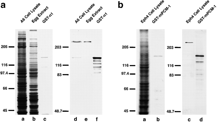
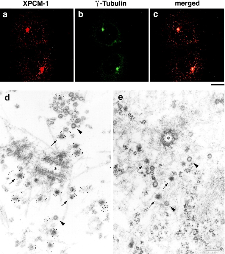
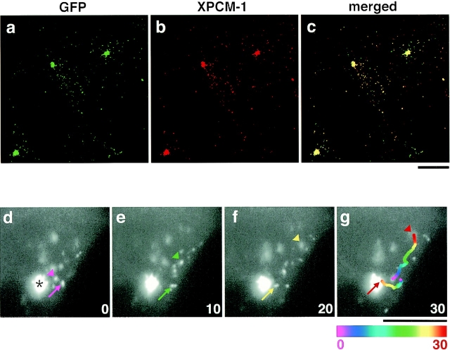
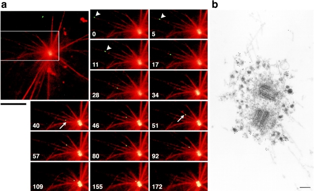
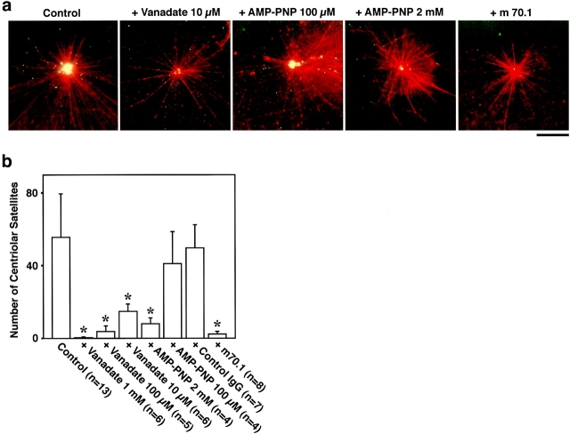
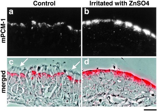
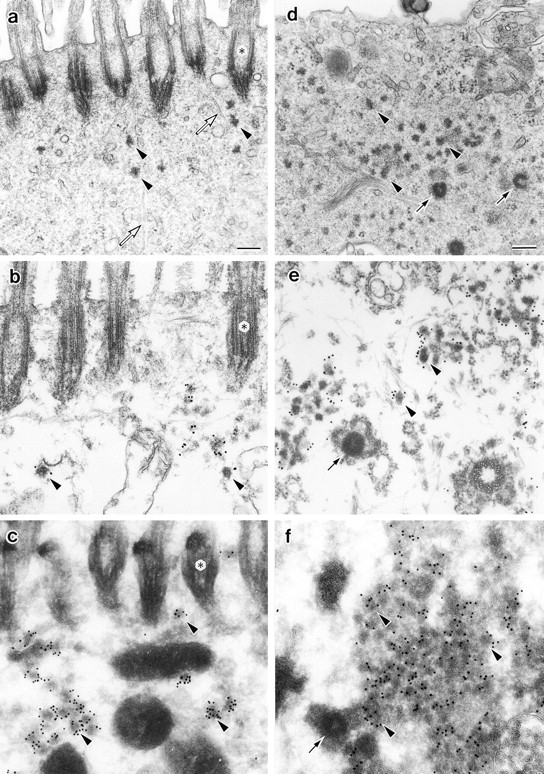
We identified Xenopus pericentriolar material-1 (PCM-1), which had been reported to constitute pericentriolar material, cloned its cDNA, and generated a specific pAb against this molecule. Immunolabeling revealed that PCM-1 was not a pericentriolar material protein, but a specific component of centriolar satellites, morphologically characterized as electron-dense granules, approximately 70-100 nm in diameter, scattered around centrosomes. Using a GFP fusion protein with PCM-1, we found that PCM-1-containing centriolar satellites moved along microtubules toward their minus ends, i.e., toward centrosomes, in live cells, as well as in vitro reconstituted asters. These findings defined centriolar satellites at the molecular level, and explained their pericentriolar localization. Next, to understand the relationship between centriolar satellites and centriolar replication, we examined the expression and subcellular localization of PCM-1 in ciliated epithelial cells during ciliogenesis. When ciliogenesis was induced in mouse nasal respiratory epithelial cells, PCM-1 immunofluorescence was markedly elevated at the apical cytoplasm. At the electron microscopic level, anti-PCM-1 pAb exclusively labeled fibrous granules, but not deuterosomes, both of which have been suggested to play central roles in centriolar replication in ciliogenesis. These findings suggested that centriolar satellites and fibrous granules are identical novel nonmembranous organelles containing PCM-1, which may play some important role(s) in centriolar replication.


Figures

Figure 1
Figure 1
Specificity of pAbs. a, Rabbit anti–Xenopus PCM-1 (XPCM-1) pAb. Total cell lysate of cultured Xenopus A6 cells (A6 Cell Lysate) and Xenopus egg extract (Egg Extract), and purified GST fusion protein with the COOH-terminal portion of XPCM-1 produced in E. coli (GST-n1) were separated by SDS-PAGE (a–c; CBB staining), followed by immunoblotting with anti–XPCM-1 pAb (d–f). This affinity-purified pAb specifically recognized ∼230 kD XPCM-1 in d and e, as well as GST fusion protein in f. b, Rabbit anti–mouse PCM-1 (mPCM-1) pAb. Total cell lysate of cultured mouse Eph4 cells (Eph4 Cell Lysate) and purified GST fusion protein with the COOH-terminal portion of mPCM-1 produced in E. coli (GST-mPCM-1) were separated by SDS-PAGE (a and b; CBB staining), followed by immunoblotting with anti–mPCM-1 pAb (c and d). This affinity-purified pAb specifically recognized ∼230 kD mPCM-1 in c, as well as GST fusion protein in d.
Figure 2
Figure 2
Subcellular localization of XPCM-1 in A6 cells. a–c, Double immunofluorescence staining of A6 cells with anti–XPCM-1 pAb (a) and anti–γ-tubulin mAb (b). The merged image (c) revealed that most of the XPCM-1–positive granular structures were concentrated on and/or around γ-tubulin–positive centrosomes. Small numbers of XPCM-1–positive granular structures were observed scattered in the cytoplasm. Bar, 10 μm. d, Localization of XPCM-1 at centriolar satellites. When A6 cells were treated with Triton X-100 and labeled with anti–XPCM-1 pAb, numerous electron dense granules (arrows) around the centrosome (asterisk) were specifically labeled at the electron microscopic level. Some granules appeared to be associated with MTs. Note that pale granules (arrowheads) with similar diameter were not labeled. e, Ultrastructure of centrosomes of A6 cells in situ. A6 cells were processed for ultrathin EM without Triton X-100 treatment. Note the so-called centriolar satellites (arrows) and pale granules (arrowheads) around the centrosome (asterisk). Bar, 200 nm.
Figure 3
Figure 3
Movement of GFP-tagged centriolar satellites in live A6 cells. a–c, GMX-A6 cells, A6 transfectants stably expressing GFP fusion protein with the middle portion of XPCM-1 (GFP-MX), were stained red with anti–XPCM-1 pAb. Since this pAb did not recognize GFP-MX, it represented the localization of endogenous XPCM-1 (b). Most of the green fluorescence from expressed GFP-MX (a) was colocalized with endogenous XPCM-1 (b and c). d–g, Image series of GMX-A6 cells showing the movement of GFP-tagged centriolar satellites. Numbers at the bottom right indicate the time lapse in seconds. Arrows show the movement of granules toward the centrosome (asterisk), and arrowheads indicate granules moving toward the cell periphery. When the granules moved quickly during 1-s exposure of the CCD camera, they appeared tube-like in shape (arrow in f). In g, the paths of movement of two granules during 30 s were traced, which were represented in 2-s intervals beginning with the coolest colors (purple) and proceeding to the hottest colors (red). Bars, 10 μm.
Figure 4
Figure 4
In vitro motility of GFP-tagged centriolar satellites. a, Time-lapse observation of the movement of a centriolar satellite (green) in a reconstituted aster (red). The boxed area in the large panel is magnified in the small panels. Numbers at the bottom left in the small panels indicate the time lapse in seconds. GFP-tagged centriolar satellite (arrowheads) moved along a MT toward centrosomes where other granules had already accumulated. On the way to the centrosome, this granule changed MTs (arrows), and finally reached the centrosome. Bar, 10 μm. A QuickTime movie is available at http://www.jcb.org/cgi/content/147/5/969/F4/DC1. b, Immunoelectron microscopy of centriolar satellites accumulating around centrosomes in the in vitro reconstituted asters. Electron-dense granular structures, ∼80–90 nm in diameter, which were specifically labeled with anti–XPCM-1 pAb (10-nm gold particles), were accumulated around fibrous materials of centrosomes. These XPCM-1–containing granules were not delineated by membranes. Bar, 200 nm.
Figure 5
Figure 5
Inhibition of the accumulation of GFP-tagged centriolar satellites around centrosomes in vitro. A reconstituted aster (red) was incubated with GFP-tagged centriolar satellites (green) under the same condition as Fig. 4 a. Without additional reagents, numerous granules were accumulated around the centrosome during 10-min incubation (Control). AMP-PNP at 2 mM, but not 100 μM, significantly suppressed the accumulation of granules. 10 μM vanadate, as well as antidynein intermediate chain mAb (m70.1), also completely suppressed the accumulation. When the accumulation was suppressed, the movement of individual granules itself was always affected. Bar, 10 μm. b, The number of centriolar satellites, which were accumulated around centrosomes during 10-min incubation, were counted per individual centrosomes. Asterisks, F-test showed significant inhibition (P < 0.001).
Figure 6
Figure 6
Subcellular distribution of mouse PCM-1 (mPCM-1) in mouse nasal respiratory epithelium. When cryosections ∼0.5-μm thick of ciliated epithelium were immunofluorescently stained with anti–mPCM-1 pAb (a, red in c), mPCM-1 signal was detected at the apical cytoplasm of epithelial cells (c; a composite with the phase-contrast image). Arrows, cilia. At four days after irritation of the nasal epithelia with 1% aqueous ZnSO4 in situ, cilia were completely removed from their apical surface, and the PCM-1 signal at the apical cytoplasm was markedly elevated (b, red in d). d, a composite with the phase-contrast image. Bar, 10 μm.
Figure 7
Figure 7
Localization of mPCM-1 in nasal respiratory epithelial cells at four days after exposure to distilled water (a–c) or irritation with 1% aqueous ZnSO4 (d–f). a, Conventional ultrathin EM. Electron-dense spherical granules (arrowheads), ∼70–100 nm in diameter, which were morphologically indistinguishable from centriolar satellites, were scattered close to ciliary basal bodies (asterisks). Open arrows, microtubules. b, Preembedding immunoelectron microscopy. Nasal epithelial tissues were treated with 0.5% Triton X-100, fixed with glutaraldehyde, then labeled with anti–mPCM-1 pAb. The centriolar satellite-like granules were specifically labeled (arrowheads). c, Postembedding immunoelectron microscopy. Ultrathin cryosections of nasal epithelial cells were labeled with anti–mPCM-1 pAb. The centriolar satellite-like granules were specifically labeled (arrowheads). d, Conventional ultrathin EM. Cilia were completely removed, and at the apical cytoplasm numerous fibrous granules (arrowheads), as well as deuterosomes (arrows), appeared. e, Preembedding immunoelectron microscopy. Samples were treated with 0.5% Triton X-100, fixed with glutaraldehyde, then labeled with anti–mPCM-1 pAb. Fibrous granules (arrowheads), but not deuterosomes (arrow), were heavily labeled. Both centriolar and acentriolar pathways for centriolar replication were observed (see details in the text). f, Postembedding immunoelectron microscopy. Ultrathin cryosections were labeled with anti–mPCM-1 pAb. Fibrous granules (arrowheads), but not deuterosomes (arrow), were specifically labeled. Bars, 200 nm.

Similar articles

Cited by

References

    1. Anderson R.G.W., Brenner R.M. The formation of basal bodies (centrioles) in the rhesus monkey oviduct. J. Cell Biol. 1971;50:10–34. - PMC - PubMed
    1. Bacallao R., Antony C., Dotti C., Karsenti E., Stelzer E.H., Simons K. The subcellular organization of Madin-Darby canine kidney cells during the formation of a polarized epithelium. J. Cell Biol. 1989;109:2817–2832. - PMC - PubMed
    1. Balczon R., Bao L., Zimmer W.E. PCM-1, A 228-kD centrosome autoantigen with a distinct cell cycle distribution. J. Cell Biol. 1994;124:783–793. - PMC - PubMed
    1. Balczon R., Bao L., Zimmer W.E., Brown K., Zinkowski R.P., Brinkley B.R. Dissociation of centrosome replication events from cycles of DNA synthesis and mitotic division in hydroxyurea-arrested Chinese hamster ovary cells. J. Cell Biol. 1995;130:105–115. - PMC - PubMed
    1. Balczon R., Varden C.E., Schroer T.A. Role for microtubules in centrosome doubling in Chinese hamster ovary cells. Cell Motil. Cytoskel. 1999;42:60–72. - PubMed

MeSH terms

Associated data