. 1999 Nov 29;147(5):1073-84.
doi: 10.1083/jcb.147.5.1073.

Enhancement of endothelial cell migration and in vitro tube formation by TAP20, a novel beta 5 integrin-modulating, PKC theta-dependent protein

Affiliations

Enhancement of endothelial cell migration and in vitro tube formation by TAP20, a novel beta 5 integrin-modulating, PKC theta-dependent protein

S Tang et al. J Cell Biol. .

Abstract

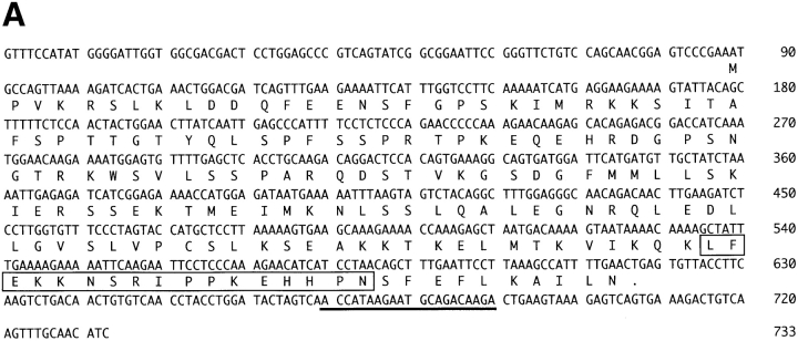
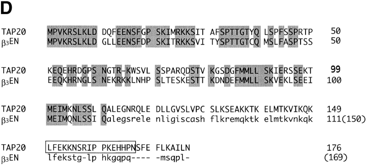
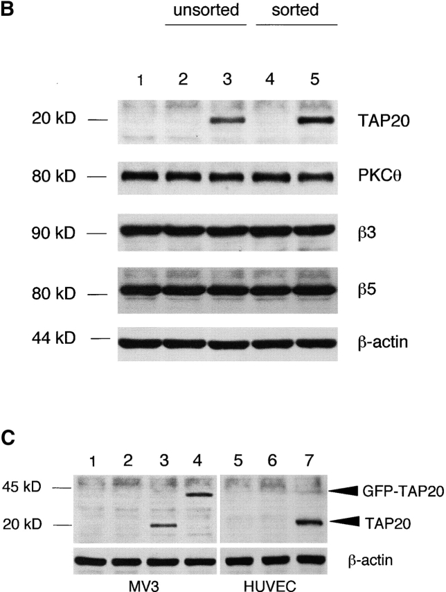
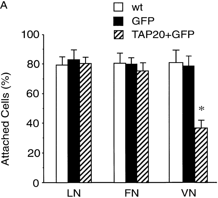
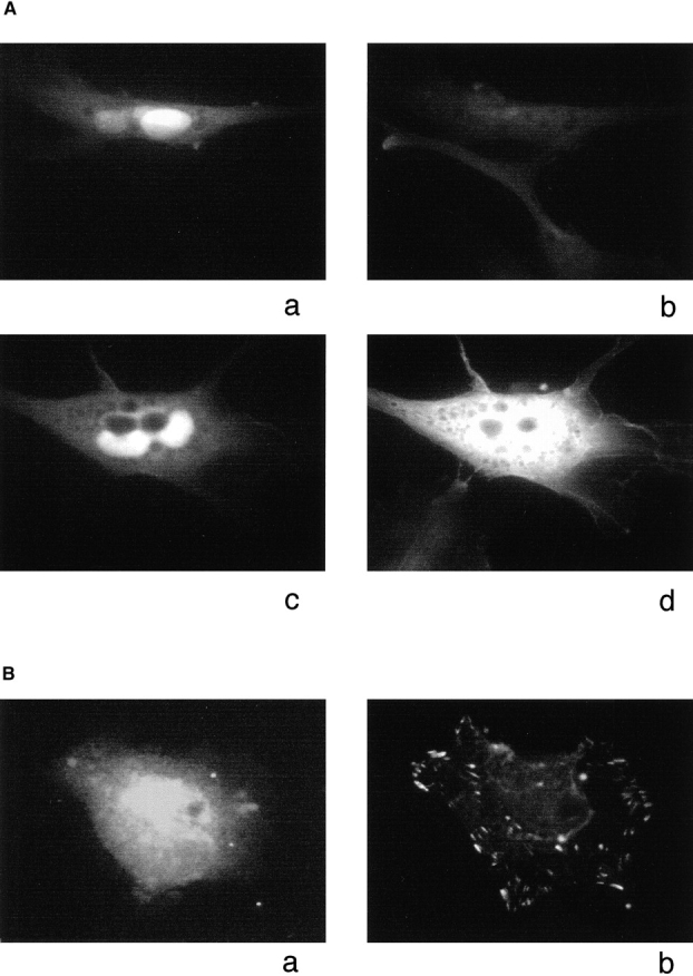
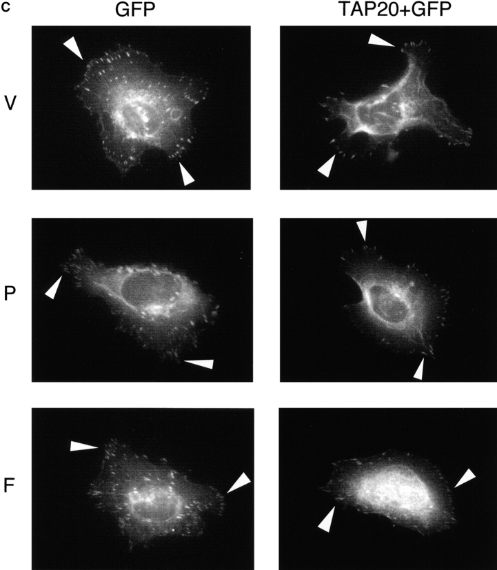
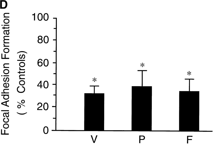
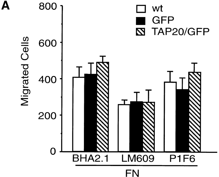
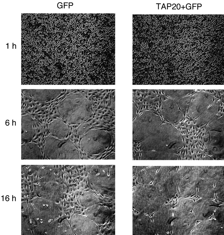
Migration, proliferation, and tube formation of endothelial cells are regulated by a protein kinase C isoenzyme PKCtheta. A full-length cDNA encoding a novel 20-kD protein, whose expression was PKCtheta-dependent, was identified in endothelial cells, cloned, characterized, and designated as theta-associated protein (TAP) 20. Overexpression of TAP20 decreased cell adhesion and enhanced migration on vitronectin and tube formation in three-dimensional culture. An antiintegrin alphavbeta5 antibody prevented these TAP20 effects. Overexpression of TAP20 also decreased focal adhesion formation in alphavbeta3-deficient cells. The interaction between TAP20 and beta5 integrin cytoplasmic domain was demonstrated by protein coprecipitation and immunoblotting. Thus, the discovery of TAP20, which interacts with integrin beta5 and modulates cell adhesion, migration, and tube formation, further defines a possible pathway to angiogenesis dependent on PKCtheta.


Figures

Figure 1
Figure 1
Molecular cloning and analysis of TAP20. (A) Nucleotide and deduced amino acid sequence of TAP20. The deduced amino acid sequence (single letter code) is shown under the nucleotide sequence. The solid line indicates the sequence used as the reverse primer to screen the rat PC12 library. The box indicates the sequence of peptide used as an immunogen to induce TAP20 antibody formation. (B and C) Differential expression of TAP20 in RCE with varying PKCθ activity levels. Total RNA (B) and RCE lysate (C) were prepared from wt RCE (wt) or cells stably expressing control plasmid (v), PKCθ-ca (ca1 and ca2), or PKCθ-kn (kn1 and kn2). The full-length TAP20 cDNA was used as probe on Northern blot (B), and the TAP20 antibody was used for Western analysis (C). (D) Amino acid sequence comparison of TAP20 with β3-endonexin. Identical amino acids are shown by the letters in gray boxes. Lower case letters indicate additional 59 amino acids of the longer form of β3-endonexin, and the boxed sequence indicates the immunogen peptide for TAP20.
Figure 1
Figure 1
Molecular cloning and analysis of TAP20. (A) Nucleotide and deduced amino acid sequence of TAP20. The deduced amino acid sequence (single letter code) is shown under the nucleotide sequence. The solid line indicates the sequence used as the reverse primer to screen the rat PC12 library. The box indicates the sequence of peptide used as an immunogen to induce TAP20 antibody formation. (B and C) Differential expression of TAP20 in RCE with varying PKCθ activity levels. Total RNA (B) and RCE lysate (C) were prepared from wt RCE (wt) or cells stably expressing control plasmid (v), PKCθ-ca (ca1 and ca2), or PKCθ-kn (kn1 and kn2). The full-length TAP20 cDNA was used as probe on Northern blot (B), and the TAP20 antibody was used for Western analysis (C). (D) Amino acid sequence comparison of TAP20 with β3-endonexin. Identical amino acids are shown by the letters in gray boxes. Lower case letters indicate additional 59 amino acids of the longer form of β3-endonexin, and the boxed sequence indicates the immunogen peptide for TAP20.
Figure 2
Figure 2
Expression of TAP20 in human cell lines. (A) Plasmid constructs. The EGFP gene with its 5′ end CMV promoter to 3′ end polyA signal region was inserted into pRc/CMV to create a GFP/pRc plasmid (GFP). TAP20 cDNA then was cloned into the multicloning site in the GFP/pRc vector. Thus, TAP20 and EGFP cDNAs were controlled by separate CMV promoters. This construct is designated as TAP20 + GFP. TAP20 cDNA was also cloned into plasmid pEGFP-C1 for expression of a GFP-TAP20 fusion protein in mammalian cells, and designated as GFP-TAP20. (B) Expression of TAP20 in human ECV cells. 1 d after transfection, cells were sorted by GFP fluorescence using FACS®. Cells with GFP expression were recultured for 1–2 d before preparing cell lysates. The lysate of unsorted cells was prepared 2–3 d after transfection. Anti-TAP20, anti-PKCθ, anti-β3, and anti-β5 antibodies were used to detect protein expression. Lane 1, wt untransfected cells (wt); lanes 2 and 4, GFP/pRc transfected cells (GFP); lanes 3 and 5, TAP20 + GFP/pRc transfected cells (TAP20 + GFP). (C) Expression of TAP20 or fusion protein GFP-TAP20 in human MV3, and expression of TAP20 in HUVEC cells. Lysates of transfected MV3 and sorted HUVEC cells were prepared as described above for immunoblotting, and anti-TAP20 antibody was used to detect protein expression. Lane 1 and 5, wt untransfected cells (wt); lanes 2 and 6, GFP/pRc transfected cells (GFP); lanes 3 and 7, TAP20 + GFP/pRc transfected cells (TAP20 + GFP); and lane 4, GFP-TAP20/pRc transfected cells (GFP-TAP20).
Figure 2
Figure 2
Expression of TAP20 in human cell lines. (A) Plasmid constructs. The EGFP gene with its 5′ end CMV promoter to 3′ end polyA signal region was inserted into pRc/CMV to create a GFP/pRc plasmid (GFP). TAP20 cDNA then was cloned into the multicloning site in the GFP/pRc vector. Thus, TAP20 and EGFP cDNAs were controlled by separate CMV promoters. This construct is designated as TAP20 + GFP. TAP20 cDNA was also cloned into plasmid pEGFP-C1 for expression of a GFP-TAP20 fusion protein in mammalian cells, and designated as GFP-TAP20. (B) Expression of TAP20 in human ECV cells. 1 d after transfection, cells were sorted by GFP fluorescence using FACS®. Cells with GFP expression were recultured for 1–2 d before preparing cell lysates. The lysate of unsorted cells was prepared 2–3 d after transfection. Anti-TAP20, anti-PKCθ, anti-β3, and anti-β5 antibodies were used to detect protein expression. Lane 1, wt untransfected cells (wt); lanes 2 and 4, GFP/pRc transfected cells (GFP); lanes 3 and 5, TAP20 + GFP/pRc transfected cells (TAP20 + GFP). (C) Expression of TAP20 or fusion protein GFP-TAP20 in human MV3, and expression of TAP20 in HUVEC cells. Lysates of transfected MV3 and sorted HUVEC cells were prepared as described above for immunoblotting, and anti-TAP20 antibody was used to detect protein expression. Lane 1 and 5, wt untransfected cells (wt); lanes 2 and 6, GFP/pRc transfected cells (GFP); lanes 3 and 7, TAP20 + GFP/pRc transfected cells (TAP20 + GFP); and lane 4, GFP-TAP20/pRc transfected cells (GFP-TAP20).
Figure 3
Figure 3
Inhibition of cell adhesion by TAP20. Transfected cells were sorted with GFP fluorescence and recultured for 1–2 d. Cells were briefly treated with trypsin, washed with PBS, and resuspended in M199 medium with 2% BSA. Cells (50,000/well) were allowed to adhere to a 96-well plate coated with LN, FN, or VN (A). Cells attached to VN were further investigated with monoclonal antiintegrin antibodies (10 μg/ml) as indicated: the anti-αvβ3 antibody, LM609; anti-αvβ5, P1F6, and as a control for integrin ligation, an anti–α2β1 integrin antibody, BHA2.1 (B). The attached EC were quantified by the dye staining method. Attached cells are expressed as the percentage of the seeded cells. The values are means from four separate experiments. Error bars indicate standard deviation; asterisk indicates P < 0.01, TAP20 transfected ECV (TAP20 + GFP) versus either wt or GFP alone transfectants, by t test.
Figure 3
Figure 3
Inhibition of cell adhesion by TAP20. Transfected cells were sorted with GFP fluorescence and recultured for 1–2 d. Cells were briefly treated with trypsin, washed with PBS, and resuspended in M199 medium with 2% BSA. Cells (50,000/well) were allowed to adhere to a 96-well plate coated with LN, FN, or VN (A). Cells attached to VN were further investigated with monoclonal antiintegrin antibodies (10 μg/ml) as indicated: the anti-αvβ3 antibody, LM609; anti-αvβ5, P1F6, and as a control for integrin ligation, an anti–α2β1 integrin antibody, BHA2.1 (B). The attached EC were quantified by the dye staining method. Attached cells are expressed as the percentage of the seeded cells. The values are means from four separate experiments. Error bars indicate standard deviation; asterisk indicates P < 0.01, TAP20 transfected ECV (TAP20 + GFP) versus either wt or GFP alone transfectants, by t test.
Figure 4
Figure 4
TAP20 modulation of focal adhesion formation on MV3 cells. 24 h after transfection with the plasmids encoding GFP, TAP20 + GFP, or the fusion protein GFP-TAP20, respectively, human MV3 cells were seeded on 4-well glass chamber slides coated with VN (10 μg/ml) and incubated in a 37°C incubator for 16 h. After fixation, cells were immunostained with antibodies. (A) Cells transfected with GFP (panels a and b) or TAP20 + GFP (panels c and d) were stained with anti-TAP20 antibody. Cells were visualized with GFP fluorescence (panels a and c) and Cy-3 fluorescence (panels b and d). Cells expressing TAP20 were strongly stained. The weak stain in TAP20-negative cells may be caused by nonspecific staining by the antibody. (B) Cells expressing the GFP-TAP20 fusion protein were visualized with GFP fluorescence (panel a) or were stained with antivinculin antibody and visualized with Cy-3 fluorescence (panel b). (C) Cells expressing GFP (left panels) or TAP20 + GFP (right panels) were stained with antibodies against vinculin (V), paxillin (P), or FAK (F). Arrowheads indicate focal adhesions. (D) Focal adhesions were quantified by counting four cells for each staining. A relative value was determined in which the number of focal adhesions in TAP20 + GFP cells is expressed as a percentage of those in control (GFP alone) cells, which were normalized at 100%. Data are expressed as the mean ± S.D. (error bars). Asterisk indicates P < 0.01, TAP20 + GFP transfectants (TAP20 + GFP) versus GFP alone transfected cells by t test.
Figure 4
Figure 4
TAP20 modulation of focal adhesion formation on MV3 cells. 24 h after transfection with the plasmids encoding GFP, TAP20 + GFP, or the fusion protein GFP-TAP20, respectively, human MV3 cells were seeded on 4-well glass chamber slides coated with VN (10 μg/ml) and incubated in a 37°C incubator for 16 h. After fixation, cells were immunostained with antibodies. (A) Cells transfected with GFP (panels a and b) or TAP20 + GFP (panels c and d) were stained with anti-TAP20 antibody. Cells were visualized with GFP fluorescence (panels a and c) and Cy-3 fluorescence (panels b and d). Cells expressing TAP20 were strongly stained. The weak stain in TAP20-negative cells may be caused by nonspecific staining by the antibody. (B) Cells expressing the GFP-TAP20 fusion protein were visualized with GFP fluorescence (panel a) or were stained with antivinculin antibody and visualized with Cy-3 fluorescence (panel b). (C) Cells expressing GFP (left panels) or TAP20 + GFP (right panels) were stained with antibodies against vinculin (V), paxillin (P), or FAK (F). Arrowheads indicate focal adhesions. (D) Focal adhesions were quantified by counting four cells for each staining. A relative value was determined in which the number of focal adhesions in TAP20 + GFP cells is expressed as a percentage of those in control (GFP alone) cells, which were normalized at 100%. Data are expressed as the mean ± S.D. (error bars). Asterisk indicates P < 0.01, TAP20 + GFP transfectants (TAP20 + GFP) versus GFP alone transfected cells by t test.
Figure 5
Figure 5
Modulation of cell migration by TAP20. Transfected HUVEC cells were sorted with GFP fluorescence and recultured for 1–2 d. Cells were briefly treated with trypsin and suspended in medium containing 0.1% BSA, with addition of antibodies as indicated: anti-α2β1, BHA2.1; anti-αvβ3, LM609; and anti-αvβ3, P1F6. Cells (50,000/0.1 ml) were seeded in coated FluoroBlok Insert precoated with either FN (A) or VN (B). Cells were incubated for 4 h at 37°C. After fixation, migrated cells were visualized under a fluorescence microscope. Cells that had migrated through the membrane of FluoroBlok Inserts were quantitated by counting the fluorescent cells in two random nonoverlapped 100× view fields. (A) Data are expressed as the mean ± S.D. (error bars) of four separate experiments. Asterisk indicates P < 0.01, TAP20 transfectants (TAP + GFP) versus GFP alone transfected cells by t test.
Figure 5
Figure 5
Modulation of cell migration by TAP20. Transfected HUVEC cells were sorted with GFP fluorescence and recultured for 1–2 d. Cells were briefly treated with trypsin and suspended in medium containing 0.1% BSA, with addition of antibodies as indicated: anti-α2β1, BHA2.1; anti-αvβ3, LM609; and anti-αvβ3, P1F6. Cells (50,000/0.1 ml) were seeded in coated FluoroBlok Insert precoated with either FN (A) or VN (B). Cells were incubated for 4 h at 37°C. After fixation, migrated cells were visualized under a fluorescence microscope. Cells that had migrated through the membrane of FluoroBlok Inserts were quantitated by counting the fluorescent cells in two random nonoverlapped 100× view fields. (A) Data are expressed as the mean ± S.D. (error bars) of four separate experiments. Asterisk indicates P < 0.01, TAP20 transfectants (TAP + GFP) versus GFP alone transfected cells by t test.
Figure 6
Figure 6
Enhancement of HUVEC tube formation on matrix gel by TAP20. Transfected cells were sorted with GFP fluorescence and were seeded on top of the matrix gel with complete M199 medium. The photographs were taken using light microscopy (100× view field) with a video TV camera system, at the timepoints indicated.
Figure 7
Figure 7
Interaction of TAP20 with β5 integrin subunit. (A) Western analysis of proteins from ECV lysate precipitated by immobilized GST (control) or GST-TAP20 fusion protein. Antibodies used for the blots are indicated. Lane 1, ECV lysate alone; lane 2, cell proteins coprecipitated with GST alone; lane 3, cell proteins coprecipitated with GST-TAP20 fusion protein. Arrowheads indicate the proteins detected. (B) Western analysis of TAP20 protein in ECV cell lysate precipitated by GST-integrin tail fusion protein, using TAP20 antibody. Lane 1, the lysate of ECV cells transfected with TAP20 + GFP; lane 2, protein precipitated by GST–β3 tail fusion protein–coated beads from the cell lysate described in lane 1; lane 3, protein precipitated by the GST–β5 tail fusion protein–coated beads from the cell lysate described in lane 1. (C) Western analysis of TAP20 protein precipitated by GST-integrin tail fusion protein, using TAP20 antibody. Lane 1, lysate from E. coli BL21 cells expressing GST-TAP20 fusion protein; lane 2, purified GST-TAP20 fusion protein digested with thrombin, precipitated by the GST–β3 cytoplasmic domain fusion protein–coated beads for 20 h; lane 3, TAP20 protein precipitated by GST–β5 cytoplasmic domain fusion protein–coated beads at 4°C for 20 h.
Figure 7
Figure 7
Interaction of TAP20 with β5 integrin subunit. (A) Western analysis of proteins from ECV lysate precipitated by immobilized GST (control) or GST-TAP20 fusion protein. Antibodies used for the blots are indicated. Lane 1, ECV lysate alone; lane 2, cell proteins coprecipitated with GST alone; lane 3, cell proteins coprecipitated with GST-TAP20 fusion protein. Arrowheads indicate the proteins detected. (B) Western analysis of TAP20 protein in ECV cell lysate precipitated by GST-integrin tail fusion protein, using TAP20 antibody. Lane 1, the lysate of ECV cells transfected with TAP20 + GFP; lane 2, protein precipitated by GST–β3 tail fusion protein–coated beads from the cell lysate described in lane 1; lane 3, protein precipitated by the GST–β5 tail fusion protein–coated beads from the cell lysate described in lane 1. (C) Western analysis of TAP20 protein precipitated by GST-integrin tail fusion protein, using TAP20 antibody. Lane 1, lysate from E. coli BL21 cells expressing GST-TAP20 fusion protein; lane 2, purified GST-TAP20 fusion protein digested with thrombin, precipitated by the GST–β3 cytoplasmic domain fusion protein–coated beads for 20 h; lane 3, TAP20 protein precipitated by GST–β5 cytoplasmic domain fusion protein–coated beads at 4°C for 20 h.

Similar articles

Cited by

References

    1. Chang D.D., Wong C., Smith H., Liu J. ICAP-1, a novel β1 integrin cytoplasmic domain-associated protein, binds to a conserved and functionally important NPXY sequence motif of β1 integrin. J. Cell Biol. 1997;138:1149–1157. - PMC - PubMed
    1. Chen Y.P., Djaffar I., Pidard D., Steiner B., Cieutat A.M., Caen J.P., Rosa J.P. Ser-752→Pro mutation in the cytoplasmic domain of integrin beta 3 subunit and defective activation of platelet integrin alpha IIb beta 3 (glycoprotein IIb-IIIa) in a variant of Glanzmann thrombasthenia. Proc. Natl. Acad. Sci. USA. 1992;89:10169–10173. - PMC - PubMed
    1. Clark E.A., Brugge J.S. Integrins and signal transduction pathwaysthe road taken. Science. 1995;268:233–239. - PubMed
    1. Clark E.A., Hynes R.O. 1997 Keystone symposium on signal transduction by cell adhesion receptors. Biochim. Biophys. Acta. 1997;1333:R9–R16. - PubMed
    1. Crowe D.T., Chiu H., Fong S., Weissman I.L. Regulation of the avidity of integrin alpha 4 beta 7 by the beta 7 cytoplasmic domain. J. Biol. Chem. 1994;269:14411–14418. - PubMed

Publication types

MeSH terms