. 1999 Dec 1;521 Pt 2(Pt 2):365-74.
doi: 10.1111/j.1469-7793.1999.00365.x.

Co-regulation of synaptic efficacy at stable polyneuronally innervated neuromuscular junctions in reinnervated rat muscle

Affiliations

Co-regulation of synaptic efficacy at stable polyneuronally innervated neuromuscular junctions in reinnervated rat muscle

E M Costanzo et al. J Physiol. .

Abstract

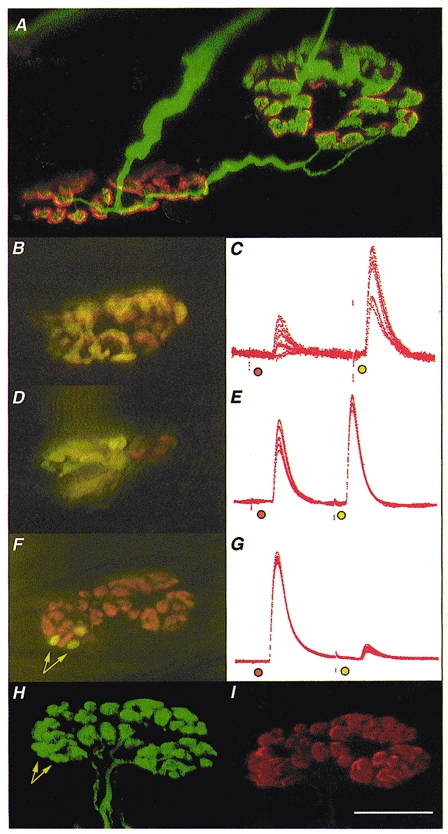
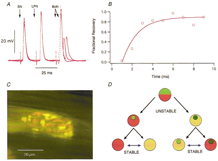
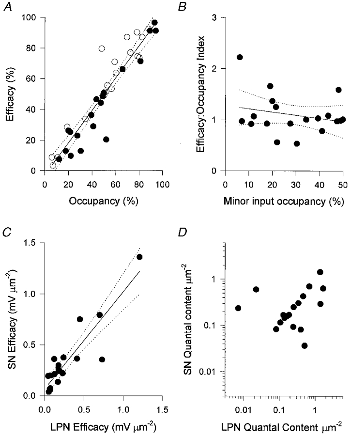
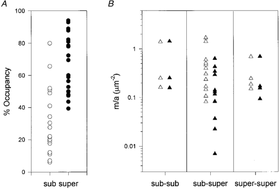
1. Intracellular recordings and quantal analysis of synaptic transmission were made at neuromuscular junctions receiving stable convergent innervation in reinnervated rat lumbrical muscles, following recovery from chronic nerve conduction block. The polyneuronally innervated motor endplates (pi-junctions) were identified by vital staining of lateral plantar nerve (LPN) and sural nerve (SN) motor terminals, using the activity-dependent staining properties of the aminostyryl dyes RH414 and FM1-43, respectively. 2. Endplate depolarisation and quantal content per unit area varied by more than a factor of ten ( approximately 0.1-1. 4 quanta microm-2) between fibres. However, the stable pi-junctions produced nearly equivalent endplate depolarisations and quantal content per unit area, suggesting that synaptic strengths were co-regulated at these motor endplates. Quantal content per unit area was also independent of the size of individual synaptic inputs, or whether one, both or neither input was judged sufficient to produce suprathreshold or subthreshold endplate depolarisations. 3. Simultaneous excitation of convergent LPN and SN inputs from some pi-junctions resulted in profound non-linear summation, and in some cases complete occlusion of the response of the smaller input. The amplitude of the smaller, test responses recovered with a time constant of 2.1 +/- 0.5 ms (mean +/- s.e.m.) on varying the interval between paired stimuli, of similar order to the time constant of repolarisation of the conditioning endplate potential. 4. The data show that it is not necessary for a motor nerve terminal to occupy most of an endplate, or to produce a suprathreshold response in order to become stable. The occlusion of linear summation, similar to that described previously at polyneuronal junctions in neonates, suggests that convergent inputs comprising interdigitated synaptic boutons evoke self-contained synaptic responses at endplates, and that these are non-co-operative with respect to overall endplate depolarisation or safety margin for synaptic transmission.


Figures

Figure 1
Figure 1. Morphology and electrophysiology of stable π-junctions
Immunocytochemistry was used to visualise nerve terminals (FITC) and TRITC-α-BTX was used to visualise AChRs in fixed preparations; FM1-43 and RH414 were used to label synaptic boutons supplied by SN and LPN motor axons in freshly isolated preparations, staining their recycled synaptic vesicles fluorescent green/yellow and orange, respectively. A, confocal microscope image of two motor endplates in a reinnervated muscle, 9 weeks after LPN crush (including 4 weeks recovery from a 2 week nerve conduction block, applied 3 weeks after the original crush). The endplates are bridged by an axonal sprout, probably arising from the motor nerve terminal on the left. The endplate on the right is also innervated by a slender regenerating axon. Thus, this endplate is a stable π-junction. B, D and F, composite digital images from three different, vitally stained stable π-junctions innervated by LPN (orange, RH414 labelled) and SN (yellow/green, FM1-43 labelled) terminal boutons, several weeks after regeneration and recovery from nerve conduction block. The image in F is a montage made up from two original digital images taken in slightly different focal planes. C, E and G, corresponding intracellular recordings of EPPs evoked from these π-junctions after bathing the preparations in μ-conotoxin to abolish muscle (but not axonal) action potentials. In each case the orange spot indicates LPN stimulation and the yellow/green spot indicates SN stimulation. The relative EPP amplitudes varied in proportion to the relative areas covered by LPN and SN synaptic boutons. The π-junction shown in F was relocated using a confocal microscope after fixing and staining for neurofilament/SV2 (H; FITC-conjugated secondary antibody, green fluorescence) and ACh receptors (I; TRITC-α-BTX, red fluorescence). Data from this endplate were as follows: in response to stimulating the nerve supplies repeatedly at 1 Hz, the LPN produced a large amplitude EPP (40 ± 0.167 mV, n = 110; uncorrected amplitudes; 93 % of the total synaptic response; mean quantal content, 47.4) and the SN produced a proportionally smaller amplitude EPP (2.9 ± 0.168 mV; 7 % of the total synaptic response; 13.8 times smaller than LPN response; mean quantal content, 3.1). The synaptic area covered by the LPN was 137 μm2 (93.2 % of total area) and the SN covered 10 μm2 (arrows in F; 6.8 % of total area; 13.7 times smaller than LPN). The confocal images confirmed that the differently coloured boutons were supplied by distinct axons directed to the same endplate, and that all boutons visualised immunocytochemically were also stained with the vital dyes before fixation. Arrows in H point to the location of the same boutons as indicated in F. There was no discernible difference in the fluorescence intensity of receptors in the region of the endplate occupied by the three SN boutons compared with receptors occupied by LPN boutons. Calibrations: scale bar in I corresponds to the following measures: A, 10 μm; B, D and F, 20 μm; H and I, 15 μm; C, E and G, 25 ms. The electrophysiological records were scaled to match the largest EPP in each case. These were as follows: C, 6 mV; E, 24 mV; G, 42 mV.
Figure 4
Figure 4. Some convergent inputs are mutually occlusive
A, three superimposed records showing EPPs evoked by SN, LPN and combined (Both) stimulation, with variable intervals between the combined stimuli in two different π-junctions. Stimulation timed to make the two peaks coincide produced no additional increment in EPP amplitude (occlusion). Assuming conventional non-linear summation of endplate conductance (McLachlan & Martin, 1981) the amplitude of the combined response should have been at least 10 mV greater than the response to LPN stimulation alone. By increasing the interval between the two stimuli, the LPN EPP was used as the conditioning input and the SN EPP as the test input. EPP occlusion was still virtually complete even when many of the AChRs were blocked, following addition of α-bungarotoxin (10 μg ml−1) to the bathing medium (not shown). B, time course of recovery from occlusion (○) and best non-linear least-squares single exponential curve fit of the form Vt = V(1 - e-t/τ) for the SN EPP shown in A. The recovery time constant was longer than the decay time constant of the conditioning EPPs. C, example of a stable π-junction where the boutons derived from the SN (green/yellow) and LPN (orange) are interdigitated. D, mononeuronal and polyneuronal innervation represented as alternative stable states, in which synaptic boutons belonging to one or more inputs may be co-regulated. Saturation of orange or green colour is used to represent synaptic strengths of two different inputs in terms of overall efficacy (EPP amplitude) and/or quantal content per unit area. The relative areas covered by orange or green, and the relative synaptic efficacies or strengths (colour saturation), may initially change due to synaptic competition and synapse elimination. Data on the effects of chronic use or disuse at mono-innervated junctions, once established, have shown that synaptic strength continues to be labile (see references in text). The present study extends this to show that, with time, stable synaptic boutons supplied by different motoneurones to the same muscle fibre become equivalent. The data further suggest that the synaptic efficacies or strengths may be scaled up or down together, i.e. they may be interconvertible (reversible blue arrows). Mismatches in the strengths of convergent inputs (light green/dark orange or dark green/light orange; upper icons) may represent a transient state characteristic of a critical period leading to synapse elimination at some junctions (left) and mononeuronal innervation as in development, or to stable polyneuronal innervation with equivalent, co-regulated synaptic strengths (light with light, or dark with dark; right) as shown by the present study.
Figure 2
Figure 2. Convergent inputs to stable π-junctions have equivalent synaptic strengths
A, EPP amplitudes corrected for non-linear summation plotted against nerve terminal area for LPN (○) and SN (•). The two measures are expressed as a percentage of the arithmetic sum of the EPP amplitudes and terminal areas, respectively. The correlation coefficient was 0.94 and regression analysis (continuous line) and 95 % confidence limits (dotted lines) showed no significant deviation from linearity over the entire range of percentage occupancies. B, efficacy:occupancy (e:o) indices calculated for the minor inputs (less than 50 % occupancy). The lack of correlation, or consistent evidence that small inputs produce EPPs smaller than others for their size suggests that even those inputs with fractional occupancies of less than 20 % of the endplate are not disproportionately weak. C, the specific efficacies of converging inputs, calculated as the EPP amplitude per unit area - corrected for non-linear summation and normalised to a resting membrane potential of -80 mV - were highly correlated (r = 0.90). This correlation could not be attributed to passive electrical properties of the muscle fibres alone, because the quantal content per unit area of the converging inputs was also highly correlated (D; r = 0.62; P < 0.005).
Figure 3
Figure 3. Suprathreshold and subthreshold synaptic inputs have similar synaptic strengths
A, the size of terminals (expressed as fractional occupancy of the endplate) required to evoke suprathreshold (super) responses in the muscle fibres was estimated assuming an action potential threshold of -63 mV from a resting potential of -75 mV (Wood & Slater, 1997). None of the inputs with fractional occupancies less than 40 % of total synaptic area were predicted to give suprathreshold responses, although some inputs occupying more than this fraction were predicted from their EPP amplitudes to give subthreshold (sub) responses. B, transmitter release per unit area (m/a) plotted for the three categories of endplate based on the capacity of their inputs to evoke suprathreshold responses: sub-sub, both inputs calculated to produce subthreshold responses; sub-super, one of the two inputs only calculated to give suprathreshold responses; super-super, both inputs calculated to be suprathreshold. In some of the sub-super fibres the subthreshold inputs showed greater and the suprathreshold inputs showed weaker synaptic strengths per unit area, but the differences were not quite statistically significant (P > 0.05, paired t test).

Similar articles

Cited by

References

    1. Attwell D, Iles JF. Synaptic transmission: ion concentration changes in the synaptic cleft. Proceedings of the Royal Society B. 1979;206:115–131. - PubMed
    1. Balice-Gordon R, Lichtman JW. Long-term synapse loss induced by focal blockade of postsynaptic receptors. Nature. 1994;372:519–524. - PubMed
    1. Barry JA, Ribchester RR. Persistent polyneuronal innervation in partially denervated rat muscle after reinnervation and recovery from prolonged nerve-conduction block. Journal of Neuroscience. 1995;15:6327–6339. - PMC - PubMed
    1. Betz WJ, Caldwell JH, Ribchester RR. The size of motor units during postnatal development of rat lumbrical muscle. The Journal of Physiology. 1979;297:463–478. - PMC - PubMed
    1. Betz WJ, Chua M, Ridge RMAP. Inhibitory interactions between motoneurone terminals in neonatal rat lumbrical muscle. The Journal of Physiology. 1989;418:25–51. - PMC - PubMed

Publication types

Substances

LinkOut - more resources