. 1999 Dec 1;521 Pt 2(Pt 2):375-88.
doi: 10.1111/j.1469-7793.1999.00375.x.

Peroxynitrite is a positive inotropic agent in atrial and ventricular fibres of the frog heart

Affiliations

Peroxynitrite is a positive inotropic agent in atrial and ventricular fibres of the frog heart

J M Chesnais et al. J Physiol. .

Abstract

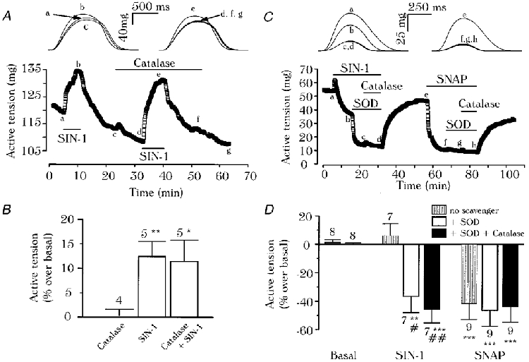
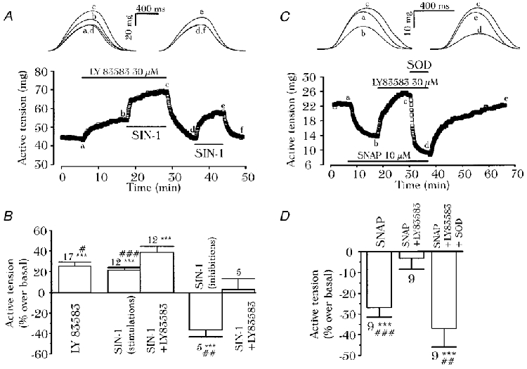
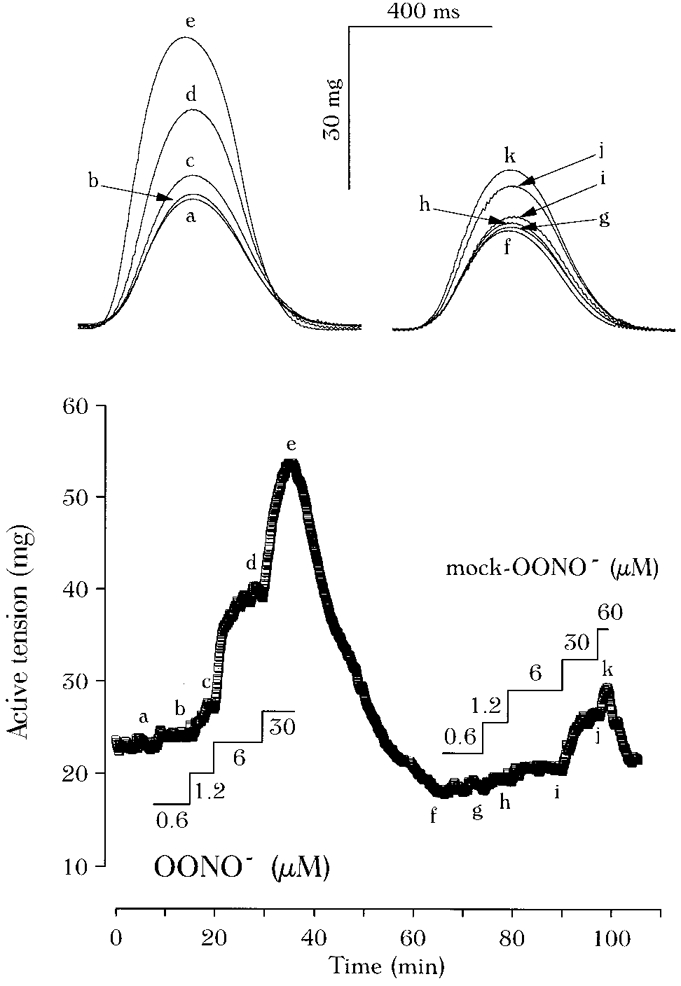
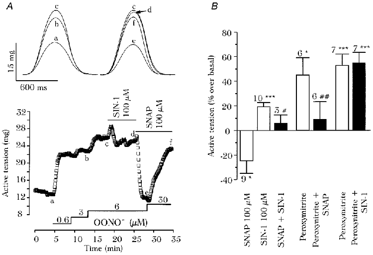
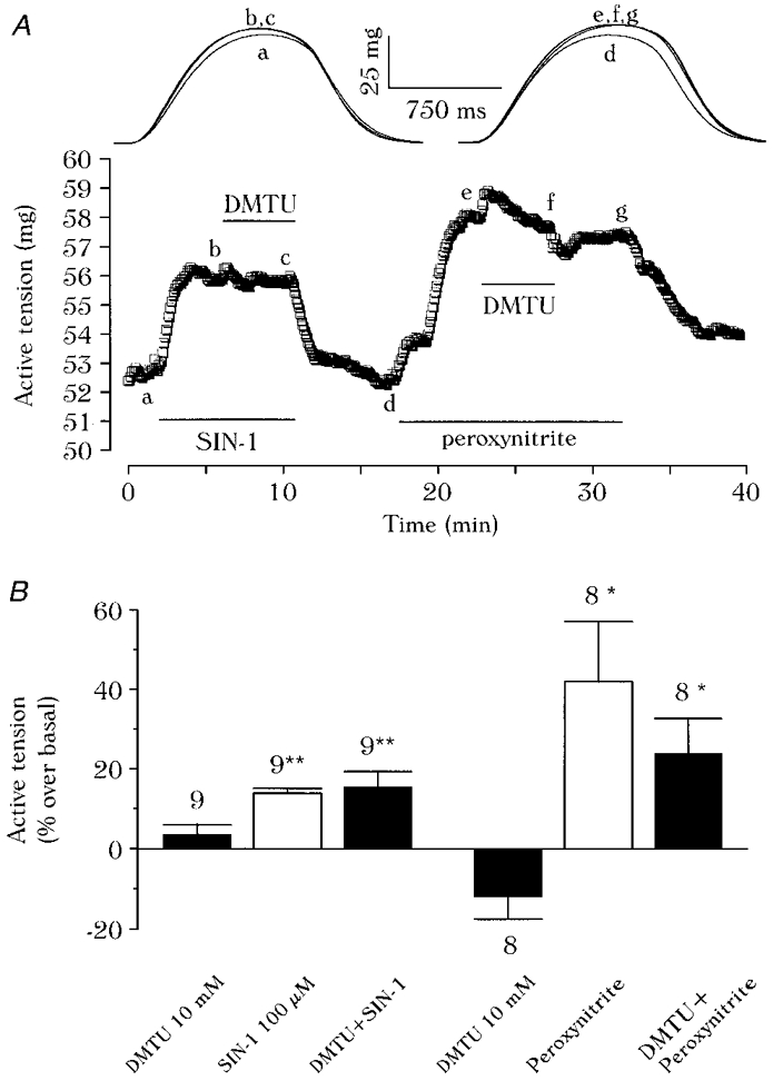
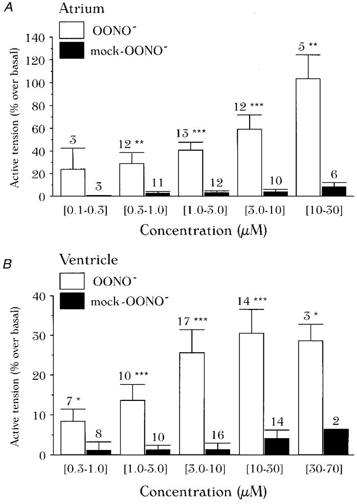
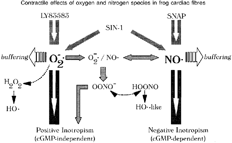
1. We report opposite inotropic effects of NO donors in frog cardiac fibres. The negative effect, elicited by either 3-morpholino-sydnonimine (SIN-1) or S-nitroso-N-acetyl-penicillamine (SNAP), involved cyclic GMP (cGMP) production. However, SIN-1, unlike SNAP, could elicit a positive effect, in a superoxide dismutase (SOD)-sensitive manner. SIN-1, unlike SNAP, can release both NO and superoxide anion, the precursors of peroxynitrite (OONO-). The role of these messengers was examined. 2. Catalase did not reduce the positive inotropic effect of SIN-1. Thus, a conversion of superoxide anion into hydrogen peroxide was not involved in this effect. In addition, catalase did not modify the negative effects of SIN-1 plus SOD, or SNAP plus SOD. 3. LY 83583, a superoxide anion generator, elicited a positive inotropic effect, like SIN-1. The effect of LY 83583 was additive to the negative effects of SIN-1 or SNAP, and to the positive effect of SIN-1. Thus, superoxide anion generation, per se, did not account for the positive effect of SIN-1. 4. Authentic peroxynitrite (OONO-), but not mock-OONO- (negative control plus decomposed OONO-), exerted a dramatic positive inotropic effect in cardiac fibres. The effect of OONO- was larger in atrial fibres, as compared with ventricular fibres. 5. The positive effect of OONO- was not additive with that of SIN-1, suggesting a common mechanism of action. In contrast, the effects of either OONO- or SIN-1 were additive with the negative inotropic effect of SNAP. Furthermore, the effect of OONO-, like that of SIN-1, was not antagonized by 1H-[1,2,4]xidiazolo[4, 3-a]quinoxaline-1-one (ODQ; 10 microM), the guanylyl cyclase inhibitor. 6. The positive inotropic effects of SIN-1 and OONO- were not modified by hydroxyl radical scavengers, such as dimethyl-thio-urea (DMTU; 10 mM). 7. The positive inotropic effect of SIN-1 (100 microM) was abolished in sodium-free solutions, a treatment that eliminates the activity of the sodium-calcium exchanger. In contrast, the effect of SIN-1 was unchanged by a potassium channel inhibitor (tetraethyl-ammonium, 20 mM), or a sodium-potassium pump inhibitor (ouabain 10 microM). 8. We conclude that OONO- is a positive inotropic agent in frog cardiac fibres. The generation of OONO- accounts for the positive inotropic effect of SIN-1. OONO- itself was responsible for the positive inotropic effect, and appeared to modulate the activity of the sodium-calcium exchanger.
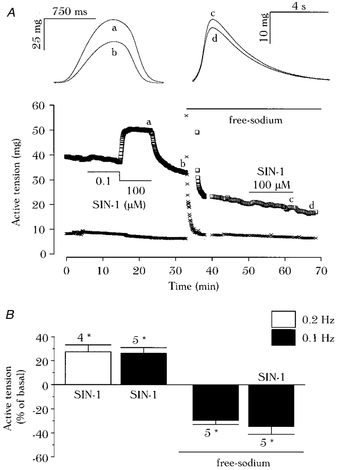

Figures

Figure 1
Figure 1. Effect of catalase in the presence of NO donors in frog cardiac fibres
A ventricular (A) or an atrial (C) fibre was initially superfused with control solution. In A, SIN-1 (100 μm) was applied in the absence or presence of catalase (1000 U ml−1), as indicated by the lines. In C, SOD (50 U ml−1) and catalase (200 U ml−1) were added in the presence of SIN-1 (100 μm) or SNAP (100 μm). Top, traces were recorded at the times indicated by the corresponding letters on the main graphs. B, summary of the effects of catalase (1000 U ml−1) and SIN-1 (100 μm), alone or in combination, in ventricular fibres where SIN-1 had induced positive effects. D, summary of the effects of SNAP (100 μm) or SIN-1 (100 μm) in the presence of SOD (50-200 U ml−1), with or without catalase (200-1000 U ml−1). In B and D, the active tension in the presence of agents was normalised to its value in the absence of agents. Bars and lines are the mean ±s.e.m. of the number of experiments indicated. Statistical differences from the basal level (*) or from SIN-1 (#) are indicated as *#P < 0.05; **##P < 0.01; ***P < 0.005.
Figure 2
Figure 2. Effects of LY 83583 and NO donors in frog cardiac fibres
The control solution was superfused onto a ventricular (A) or an atrial (C) fibre. In A, SIN-1 (100 μm) and LY 83583 (30 μm) were added alone or in combination, as indicated by the lines. In C, SNAP (10 μm), LY 83583 (30 μm), and SOD (370 U ml−1) were successively added to the solution. Top, traces were recorded at the times indicated by the corresponding letters on the main graphs. B summarises the inotropic effects of LY 83583 (10 μm) and SIN-1 (100 μm). Positive (11 ventricular plus 1 atrial fibre) and negative (5 atrial fibres) inotropic effects of SIN-1 were separated. D summarises the inotropic effects of SNAP (5-20 μm), alone, or in the presence of LY 83583 (30 μm), with or without SOD (200-370 U ml−1). The active tension in the presence of agents was normalised to its value in the absence of agents. Bars and lines are the mean ±s.e.m. of the number of experiments indicated. Statistical differences from the basal level are indicated as *P < 0.05; **P < 0.01; ***P < 0.005; and differences from SIN-1 plus LY 83583 (in B), or SNAP plus LY 83583 (in D) are indicated as #P < 0.05; ##P < 0.01; ###P < 0.005.
Figure 3
Figure 3. OONO is a positive inotropic agent in frog cardiac fibres
An atrial fibre was initially superfused with the control solution. Superfusion with authentic OONO (from 0.6 to 30 μm) or with mock-OONO (from 0.6 to 60 μm) is indicated by the lines. Top, individual contractions recorded at the times indicating by the corresponding letters on the main graph.
Figure 5
Figure 5. Interaction between OONO and NO donors in frog cardiac fibres
A, an atrial fibre was first superfused with the control solution. Superfusion with authentic OONO (0.6-30 μm), SIN-1 (100 μm) and SNAP (100 μm) are indicated by the lines. Top, contractions were recorded at the times indicated by the corresponding letters on the main graphs. B, summary of the effects of OONO (6-30 μm), SIN-1 (100 μm) and SNAP (100 μm), alone or in combination, on the active tension of cardiac fibres. The active tension in the presence of agents was normalised to its value in the absence of agents. Bars and lines are the mean ±s.e.m. of the number of experiments indicated. Statistical differences from the basal level (*) or from SNAP (#) are indicated as *#P < 0.05; ##P < 0.01; ***P < 0.005.
Figure 6
Figure 6. Effect of DMTU in the presence of SIN-1 or OONO in frog cardiac fibres
In A a ventricular fibre was initially superfused with the control solution. SIN-1 (100 μm), DMTU (10 mm) and OONO (14 μm) were applied as indicated by the lines. B, summary of the effects on the active tension of SIN-1 (100 μm) and OONO (0.6-14 μm) in the absence or presence of DMTU (10 mm). The active tension in the presence of agents was normalised to its value in the absence of agents. Bars and lines are the mean ±s.e.m. of the number of experiments indicated. Statistical differences from the control level are indicated as *P < 0.05; **P < 0.01.
Figure 7
Figure 7. Effect of SIN-1 in sodium-free solution in frog cardiac fibres
In A, the amplitude of active (□) and resting (×) tensions of a ventricular fibre are shown. The fibre was superfused with SIN-1 (0.1 and/or 100 μm) in the presence and absence of sodium ions, as indicated by the lines. Top, contractions were recorded at the times indicated by the corresponding letters on the main graphs. B, summary of the effects of SIN-1 (100 μm) on the active tension in control and sodium-free solutions, elicited at 0.2 or 0.1 Hz, as indicated. Active tensions in the different conditions were normalised to the amplitude of the active tension elicited with the routine protocol (control solution, 0.2 Hz stimulation). Bars and lines are the mean ±s.e.m. of the number of experiments indicated. Statistical differences from the basal level are indicated as *P < 0.005.
Figure 4
Figure 4. Summary of the inotropic effects of OONO in frog cardiac fibres
A and B, mean effects of OONO and of mock-OONO in ventricular and atrial fibres, respectively. Individual results were pooled and averaged within the ranges of concentrations indicated. The active tension in the presence of agents was normalised to its value in the absence of agent. Bars and lines are the mean ±s.e.m. of the number of experiments indicated. Statistical differences from the basal level are indicated as *P < 0.05; **P < 0.01; ***P < 0.005.
Figure 8
Figure 8. A schematic diagram of the regulation of cardiac contractility by NO in frog heart
This illustration is discussed in detail in the Discussion section. Filled and open arrows represent the pathways that have received experimental evidence in frog cardiac fibres, dotted arrows represent reactions and pathways that have been discarded, based on the present study. NO, nitric oxide; O2·, superoxide anion; H2O2, hydrogen peroxide; HO·, hydroxyl radical; OONO, peroxynitrite (adapted from Beckman & Koppenol, 1996; Mayer et al. 1998).

References

    1. Abi Gerges N, Hove-Madsen L, Fischmeister R, Méry P-F. A comparative study of the effects of three guanylyl cyclase inhibitors on the L-type Ca2+ and muscarinic K+ currents in frog cardiac myocytes. British Journal of Pharmacology. 1997;121:1369–1377. - PMC - PubMed
    1. Beckman JS, Koppenol WH. Nitric oxide, superoxide and peroxynitrite: the good, the bad, and the ugly. American Journal of Physiology. 1996;271:C1424–1437. - PubMed
    1. Brutsaert DL, Andries LJ. The endocardial endothelium. American Journal of Physiology. 1992;263:H985–1002. - PubMed
    1. Campbell DL, Stamler JS, Strauss HC. Redox modulation of L-type calcium channels in ferret ventricular myocytes. Journal of General Physiology. 1996;108:277–293. - PMC - PubMed
    1. Chapman RL. A study of the contractures induced in frog atrial trabeculae by a reduction of the bathing sodium concentration. The Journal of Physiology. 1974;237:295–313. - PMC - PubMed

Publication types

MeSH terms

LinkOut - more resources