. 1999 Nov;98(3):363-70.
doi: 10.1046/j.1365-2567.1999.00864.x.

Nitric oxide and macrophage antiviral extrinsic activity

Affiliations

Nitric oxide and macrophage antiviral extrinsic activity

F Benencia et al. Immunology. 1999 Nov.

Abstract

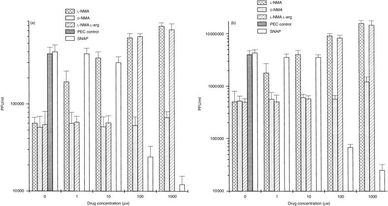
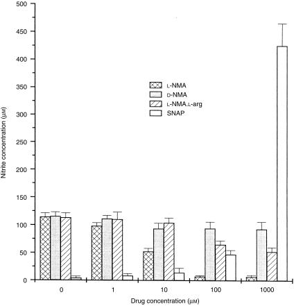
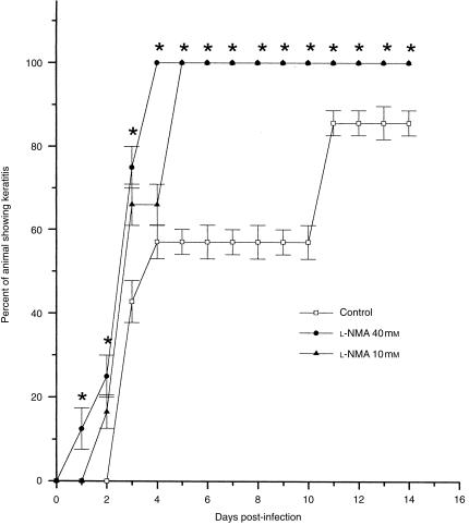
In this study we evaluated the relationship between nitric oxide (NO) and macrophage antiviral extrinsic activity. Macrophages activated by intraperitoneal injection of herpes simplex virus-2 (HSV-2), showed both extrinsic antiviral activity and high nitrite production in contrast to non-activated, resident macrophages. The extrinsic antiviral activity was observed in cultures of Vero cells infected with HSV-1 and HSV-2. The NO inhibitor N-monomethyl-l-arginine acetate (l-NMA) impaired the antiviral activity of HSV-elicited macrophages. The effect was dose dependent and correlated with a reduction of nitrite in the culture media. The effect of l-NMA was reversed by the addition of l-arginine. These data indicate that NO could be responsible for the described activity. Furthermore, l-NMA treatment resulted in the aggravation of HSV-1-induced keratitis in the mouse model, supporting a defensive role of NO in the pathogenesis of HSV-1 corneal infection.


Figures

Figure 1
Figure 1
Effect of N-monomethyl-l-arginine acetate (l-NMA) on herpes simplex virus (HSV)-elicited macrophage antiviral extrinsic activity. HSV-elicited macrophages were obtained from HSV-2-infected mice. Vero cells were grown to confluence in 24-well plates and infected with HSV-2 (a) or HSV-1 (b) before adding HSV-elicited macrophages. In peritoneal exudate cell (PEC) controls, the co-cultures were performed with non-elicited (resident) macrophages. S-nitroso-N-acetylpenicillamine (SNAP) experiments were performed using PEC controls. The plates were incubated for 2 days with medium supplemented with different concentrations of SNAP (a nitric oxide [NO] donor), l-NMA (a nitric oxide synthetase [NOS] inhibitor) and its analogue, d-NMA, or with different concentrations of l-NMA plus 1000 μm of l-arginine (l-arg). Then, cells were freeze–thawed three times in order to obtain virus. Virus yield was determined by the plaque-forming units method. Data represent the mean±SD of three independent experiments.
Figure 2
Figure 2
Nitrite production by herpes simplex virus (HSV)-elicited macrophages. Equal volumes of media from 48-hr cultures of HSV-elicited macrophages and HSV-2-infected Vero cells supplemented with different concentrations of N-monomethyl-l-arginine acetate (l-NMA), N-monomethyl-d-arginine acetate (d-NMA) or from peritoneal exudate cell (PEC) controls supplemented with various concentrations of S-nitroso-N-acetylpenicillamine (SNAP), were added to Griess reagent and incubated at room temperature for 10 min. The absorbance at 550 nm was measured using a microplate reader. NO2 concentration was determined using NaNO2 as a standard and double-distilled water as a blank. Background NO2 values of buffer or media were determined in each case and subtracted from the experimental values. Data represent the mean±SD of three independent experiments.
Figure 3
Figure 3
N-monomethyl-l-arginine acetate (l-NMA) inhibition of herpetic stromal keratitis (HSK). Mice were separated into three experimental groups receiving a 10-μl drop of l-NMA 40 mm (group 1) or 10 mm (group 2) in phosphate-buffered saline (PBS) (treated) or diluent (control; group 3), 24 hr and 2 hr before virus inoculation. After infection, mice received the corresponding treatment three times a day for 5 days. At different days postinfection, the cornea, iris and lids of animals were examined for signs of disease. Criteria for keratitis included stromal opacity, mydriasis, corneal neovascularization and corneal ulceration. Clinical evaluations were performed blind. Data represent the mean±SD of three independent experiments. *P<0·05.

Similar articles

Cited by

References

    1. Hayashi K, Kurata T, Morishima T, Nassery T. Analysis of the inhibitory effect of peritoneal macrophages on the spread of herpes simplex virus. Infect Immun. 1980;28:350. - PMC - PubMed
    1. Sit MF, Tenney DJ, Rothstein JL, Morahan PS. Effect of macrophage activation on resistance of mouse peritoneal macrophages to infection with herpes simplex virus types 1 and 2. J Gen Virol. 1988;69:1999. - PubMed
    1. Morahan PS, Morse S, McGeorge M. Macrophage extrinsic antiviral activity during herpes simplex virus infection. J Gen Virol. 1980;46:291. - PubMed
    1. Ellerman-Eriksen S. Autocrine secretion of interferon-α/β and tumour necrosis factor-α synergistically activates mouse macrophages after infection with herpes simplex virus type 2. J Gen Virol. 1993;74:2191. - PubMed
    1. Ellerman-Eriksen S, Sommerlund M, Mogensen SC. Differential sensitivity of macrophages from herpes simplex virus resistant and susceptible mice to respiratory burst priming by interferon-α/β. J Gen Virol. 1989;70:2139. - PubMed

MeSH terms

LinkOut - more resources