. 1999 Nov;98(3):413-21.
doi: 10.1046/j.1365-2567.1999.00888.x.

Interaction of CTLA-4 (CD152) with CD80 or CD86 inhibits human T-cell activation

Affiliations

Interaction of CTLA-4 (CD152) with CD80 or CD86 inhibits human T-cell activation

K Vandenborre et al. Immunology. 1999 Nov.

Abstract

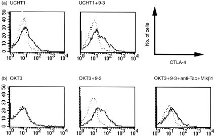
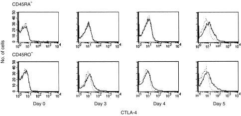
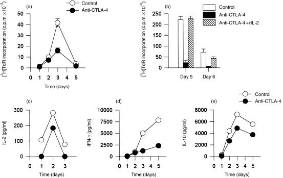
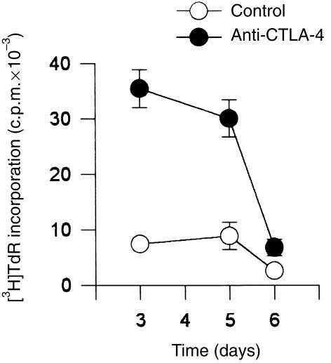
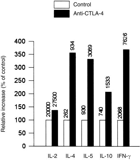
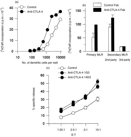
Occupancy of CTLA-4 (cytotoxic T-lymphocyte antigen-4 or CD152) negatively regulates the activation of mouse T lymphocytes, as indicated by the fate of CTLA-4-deficient mice, by the impact of anti-CTLA-4 monoclonal antibodies (mAbs) on mouse T-cell activation in vitro and by the impact of CTLA-4 blockade on the course of experimental tumoral, autoimmune, alloimmune or infectious disease in this animal. The function of human CTLA-4, however, remains less clear. The expression and function of human CTLA-4 were further explored. CTLA-4 was expressed under mitogenic conditions only, its expression being, at least partially, dependent on the secretion of interleukin-2. Memory T cells expressed CTLA-4 with faster kinetics than naive T cells. The functional role of human CTLA-4 was assessed utilizing a panel of four anti-CTLA-4 mAbs that blocked the interaction between CTLA-4 and its ligands. These mAbs, in immobilized form, profoundly inhibited the activation of T cells by immobilized anti-CD3 mAb in the absence of anti-CD28 mAb, but co-stimulated T-cell activation in the presence of anti-CD28 mAb. Finally, and importantly, blockade of the interaction of CTLA-4 with its ligands using soluble anti-CTLA-4 mAbs, in intact form or as Fab fragments, enhanced T-cell activation in several polyclonal or alloantigen-specific CD80- or CD80/CD86-dependent assays, as measured by cytokine production, cellular proliferation or cytotoxic responses. It is concluded that interaction of CTLA-4 with its functional ligands, CD80 or CD86, can down-regulate human T-cell responses, probably by intracellular signalling events and independent of CD28 occupancy.


Figures

Figure 1
Figure 1
Cytotoxic T‐lymphocyte antigen‐4 (CTLA‐4) expression on activated human T lymphocytes is enhanced by CD28 co‐stimulation and is interleukin (IL)‐2 dependent. Purified T cells were activated for 4 days with anti‐CD3 monoclonal antibody (mAb) UCHT1 immobilized on latex beads (bead to cell ratio 1 : 2) (a), or by anti‐CD3 mAb OKT3 immobilized on 24‐well plates (b), in the presence or absence of soluble anti‐CD28 mAb 9.3 (500 ng/ml), or anti‐Tac (5 µg/ml) and Mikβ1 (5 µg/ml), as indicated. Cells were stained with fluorescein isothiocyanate (FITC)‐conjugated anti‐CD5 and with biotinylated anti‐CTLA‐4 mAb BNI3.1 (IgG2a) followed by streptavidin–phycoerythrin (PE) (solid line). Histograms of anti‐CTLA‐4 reactivity of CD5‐positive cells are shown. The results are representative of three experiments. The dotted line represents the biotinylated isotype control stained with streptavidin–PE.
Figure 2
Figure 2
Human memory T cells express cytotoxic T‐lymphocyte antigen‐4 (CTLA‐4) with more rapid kinetics than naive T cells. CD45RA+ and CD45RO+ T cells, isolated by negative immunomagnetic selection, were activated with anti‐CD3 monoclonal antibody (mAb) UCHT1 immobilized on beads. Anti‐CD28 mAb 9.3 was added to a final concentration of 100 ng/ml. After the indicated intervals, cells were stained with anti‐CD5–fluorescein isothiocyanate (FITC) and biotinylated anti‐CTLA‐4 mAb BNI3.1, followed by streptavidin–phycoerythrin (PE) (solid line). Histograms of reactivity with anti‐CTLA‐4 mAb of CD5‐positive cells are shown (solid line), while the dotted line represents the isotype control. The results are representative of four experiments.
Figure 3
Figure 3
Polyclonal activation of human T cells with immobilized anti‐CD3 monoclonal antibody (mAb) is inhibited by co‐immobilized anticytotoxic T‐lymphocyte antigen‐4 (CTLA‐4) mAb. UCHT1 (anti‐CD3 mAb) and anti‐CTLA‐4 mAbs (a, 14D3; b, 1G3; c, d and e, 2G12) were co‐immobilized onto latex beads and added to cultures of purified T cells at a bead to T‐cell ratio of 2 : 1 (solid circles or bars). Beads with co‐immobilized UCHT1 and isotype‐control mAb were used as controls (open circles or bars). Where indicated, exogenous interleukin (IL)‐2 was added at time 0 to a final concentration of 10 U/ml (b, hatched bars). Cellular proliferation of quadruplicate cultures is shown as [3H]‐thymidine ([3H]TdR) incorporation ± SD (a) (b). The concentration of IL‐2 (c), interferon‐γ (IFN‐γ) (d) and IL‐10 (e) was measured by enzyme‐linked immunosorbent assay (ELISA). The data in (a), (b), (c), (d) and (e) were obtained from independent experiments.
Figure 4
Figure 4
Co‐immobilized anti‐cytotoxic T‐lymphocyte antigen‐4 (CTLA‐4) monoclonal antibody (mAb) enhances polyclonal T‐cell proliferation in the presence of soluble anti‐CD28 mAb. T cells were stimulated with co‐immobilized anti‐CD3 mAb (OKT3) and anti‐CTLA‐4 mAb 14D3 in a bead to T‐cell ratio of 2 : 1 (solid circles) in the presence of soluble anti‐CD28 mAb 9.3 at a final concentration of 100 ng/ml. In control cultures cells were stimulated with beads coated with anti‐CD3 mAb (OKT3) and an isotype control antibody (open circles), in the presence of anti‐CD28 mAb 9.3 (100 ng/ml). [3H]‐Thymidine ([3H]TdR) incorporation is shown as means of quadruplicate cultures ± SD. The results are representative of three experiments.
Figure 5
Figure 5
Blocking anti‐cytotoxic T‐lymphocyte antigen‐4 (CTLA‐4) monoclonal antibodies (mAbs) increase cytokine production in a polyclonal CD80‐dependent T‐cell activation assay. Purified human T cells were stimulated with anti‐CD3 mAb (UCHT1, 1 µg/ml) in the presence of P815/CD80 cells in a stimulator–responder ratio of 2 : 1. Soluble anti‐CTLA‐4 mAb 2G12 was added at 5 µg/ml. Cytokine production was measured after 48 hr of culture (72 hr for interleukin [IL]‐2 production) and is presented as percentage of control. For measurement of IL‐2, humanized anti‐Tac and humanized Mikβ1 were added to block IL‐2 consumption. Anti‐IL‐4 receptor (IL‐4R) was added to block IL‐4 consumption. The absolute cytokine concentration in pg/ml is shown above the bar.
Figure 6
Figure 6
Blocking the cytotoxic T‐lymphocyte antigen‐4 (CTLA‐4)–CD80/CD86 interaction enhances T‐cell proliferation and cytotoxic responses in mixed lymphocyte response (MLR) (a) 50 × 103 purified T cells were stimulated with 7·5–10 × 103 allogeneic monocyte‐derived dendritic cells for 6 days. Anti‐CTLA‐4 mAb 2G12 was added to 10 µg/ml. [3H]‐Thymidine ([3H]TdR) incorporation is shown as means of quadruplicate cultures ± SD. The results are representative of three experiments. (b) Peripheral blood mononuclear cells (PBMC) were stimulated with irradiated allogeneic PBMC in a primary mixed lymphocyte response (MLR) in the presence of Fab fragments of anti‐CTLA‐4 mAb 14D3 or isotype Fab fragments and [3H]TdR incorporation was measured after 6 days. PBMC were also stimulated with allogeneic PBMC and then restimulated in a secondary MLR. After the primary MLR (6 days), the cells were washed and rested in complete medium. After 2 days they were restimulated for 4 days with either freshly isolated PBMC from the same donor or with a third party stimulator, the B‐cell line RPMI 8866, in the presence of Fab fragments of anti‐CTLA‐4 mAb 14D3 or isotype Fab fragments, as indicated. (c) T cells were cultured for 5 days in a primary MLR against irradiated RPMI 8866 at a stimulator–reponder ratio of 1 : 20, and restimulated in a secondary MLR in the presence of either anti‐CTLA‐4 mAb 1G3 or 14D3 or control mouse IgG. After 3 days, specific cytotoxicity was measured. Means of quadruplicate samples are shown ± SD. E : T, ratio of effector cells : target cells.

Similar articles

Cited by

References

    1. June CH, Bluestone JA, Nadler LM, Thompson CB. The B7 and CD28 receptor families. Immunol Today. 1994;15:321. - PubMed
    1. Linsley PS, Greene JL, Brady W, Bajorath J, Ledbetter JA, Peach R. Human B7 (CD80) and B7 (CD86) bind with similar avidities but distinct kinetics to CD28 and CTLA‐4 receptors. Immunity. 1994;1:793. - PubMed
    1. Tivol EA, Borriello F, Schweitzer AN, Lynch WP, Bluestone JA, Sharpe AH. Loss of CTLA‐4 leads to massive lymphoproliferation and fatal multiorgan tissue destruction, revealing a critical negative regulatory role of CTLA‐4. Immunity. 1995;3:541. - PubMed
    1. Waterhouse P, Penninger JM, Timms E, et al. Lymphoproliferative disorders with early lethality in mice deficient in Ctla‐4. Science. 1995;270:985. - PubMed
    1. Krummel MF, Allison JP. CD28 and CTLA‐4 have opposing effects on the response of T cells to stimulation. J Exp Med. 1995;182:459. - PMC - PubMed

Publication types

LinkOut - more resources