. 1999 Dec 6;190(11):1549-60.
doi: 10.1084/jem.190.11.1549.

A role for lipid rafts in B cell antigen receptor signaling and antigen targeting

Affiliations

A role for lipid rafts in B cell antigen receptor signaling and antigen targeting

P C Cheng et al. J Exp Med. .

Abstract

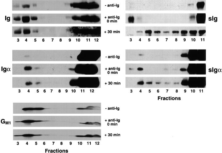
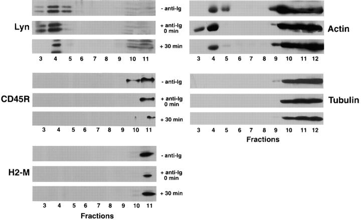
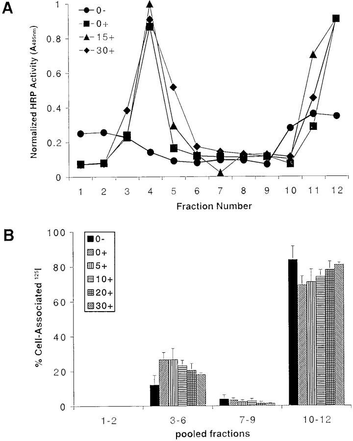
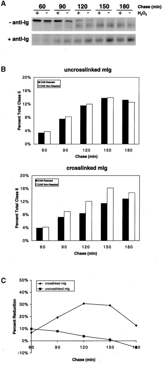
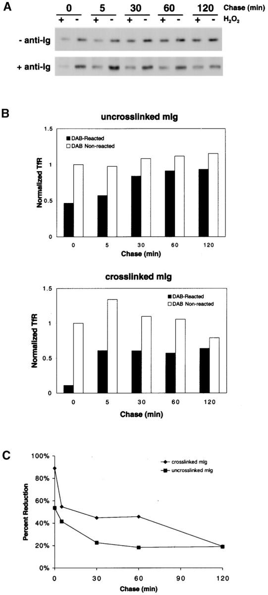
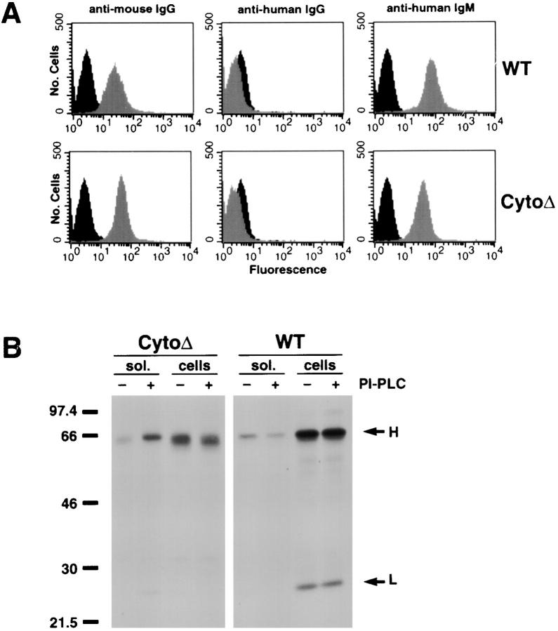
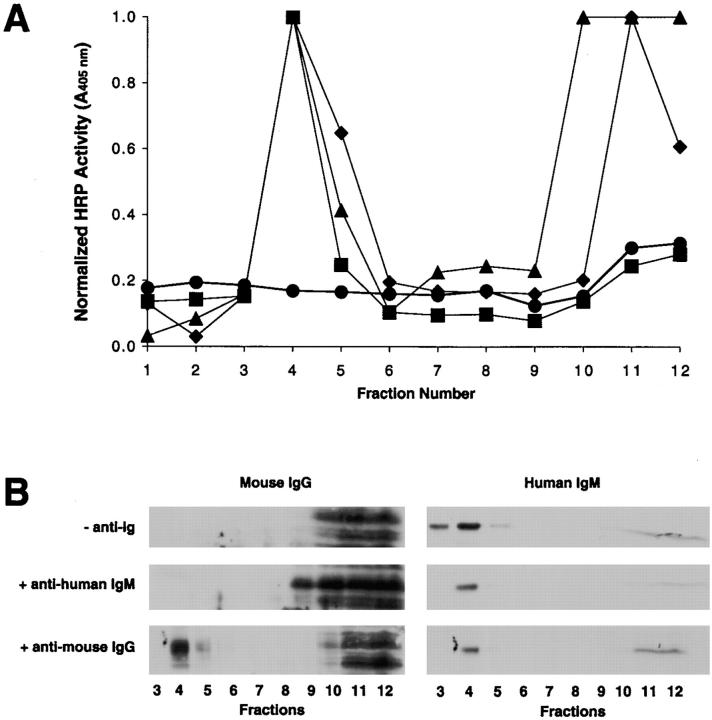
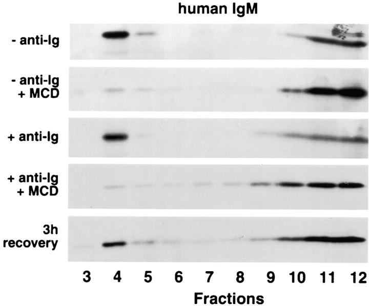
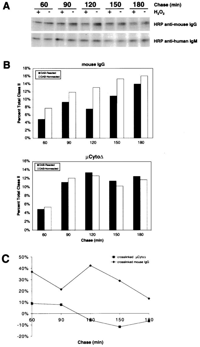
The B cell antigen receptor (BCR) serves both to initiate signal transduction cascades and to target antigen for processing and presentation by MHC class II molecules. How these two BCR functions are coordinated is not known. Recently, sphingolipid- and cholesterol-rich plasma membrane lipid microdomains, termed lipid rafts, have been identified and proposed to function as platforms for both receptor signaling and membrane trafficking. Here we show that upon cross-linking, the BCR rapidly translocates into ganglioside G(M1)-enriched lipid rafts that contain the Src family kinase Lyn and exclude the phosphatase CD45R. Both Igalpha and Lyn in the lipid rafts become phosphorylated, and subsequently the BCR and a portion of G(M1) are targeted to the class II peptide loading compartment. Entry into lipid rafts, however, is not sufficient for targeting to the antigen processing compartments, as a mutant surface Ig containing a deletion of the cytoplasmic domain is constitutively present in rafts but when cross-linked does not internalize to the antigen processing compartment. Taken together, these results provide evidence for a role for lipid rafts in the initial steps of BCR signaling and antigen targeting.


Figures

Figure 1
Figure 1
BCR cross-linking results in translocation of the BCR into GM1-containing lipid rafts. CH27 cells were surface biotinylated at 4°C and incubated with anti-Ig on ice for 15 min. The cells were warmed for 0 or 30 min and lysed in 1% Triton X-100 in TNEV buffer. The lysates were subjected to discontinuous sucrose density gradient centrifugation, and 1-ml fractions were collected. To determine the location of lipid rafts in the gradient, 100 μl of the individual fractions were subjected to 10% SDS-PAGE. After transfer onto PVDF, the membranes were probed for the presence of μ heavy chain and Igα using specific antibodies. The ganglioside GM1 was detected using CTB–HRP. To determine the location of surface BCR in the gradient, biotinylated proteins were immunoprecipitated from the gradient fractions using streptavidin–agarose. The immunoprecipitates were subjected to 10% SDS-PAGE and immunoblot probing with antibodies specific for Ig and Igα detected with HRP-conjugated secondary antibodies and ECL. Representative blots of three separate experiments are shown.
Figure 2
Figure 2
The kinase Lyn is constitutively present and the phosphatase CD45R is excluded from rafts in B cells. CH27 cells were treated with anti-Ig at 4°C for 15 min, washed, and warmed for 0 or 30 min at 37°C. The cells were lysed in 1% Triton X-100 in TNEV, and lysates were subjected to discontinuous sucrose gradient centrifugation. Fractions from the gradient were subjected to 10% SDS-PAGE and transferred to PVDF. To determine the positions in the gradient of Lyn, CD45R, H2-M, actin, and tubulin, the membranes were probed with specific antibodies followed by HRP-conjugated secondary antibodies and ECL. Shown are representative blots of three separate experiments.
Figure 3
Figure 3
Phosphorylated Igα and Lyn are present in lipid rafts after BCR cross-linking. CH27 cells were untreated or incubated with anti-Ig for 30 min at 4°C and warmed to 37°C for 0, 10, or 30 min. Cells were lysed in 1% Triton X-100 in TNEV buffer and subjected to discontinuous sucrose density gradient centrifugation. Right panels: gradient fractions were subjected to SDS-PAGE and immunoblot probing with the HRP-conjugated, phosphotyrosine-specific recombinant Ab RC20H and visualized by ECL. Left panels: immunoblots were treated with 0.02% sodium azide to inhibit RC20H enzyme activity, reprobed with Igα- and Lyn-specific mAbs, and detected using HRP-conjugated secondary antibodies and ECL. Shown are the immunoblots from fraction 4. Representative blots of three separate experiments are shown.
Figure 4
Figure 4
BCR-bound antigens translocate into rafts after BCR cross-linking. (A) B cells were untreated (0−) or treated with HRP–anti-Ig at 4°C for 1 h, washed, and warmed to 37°C for 0, 15, or 30 min (0+, 15+, and 30+, respectively). The cells were lysed in 1% Triton X-100 in TNEV buffer, the lysates subjected to discontinuous sucrose density gradient centrifugation, and the fractions assayed for HRP activity. (B) CH27 cells were incubated at 4°C for 1 h with 125I–Fab–anti-Ig. During the last 30 min of incubation, the cells were untreated (0−) or treated with anti-Ig to cross-link the BCR and then warmed to 37°C for 0–30 min (0+ to 30+). The cells were lysed in 1% Triton X-100 in TNEV buffer, the lysates were subjected to discontinuous sucrose gradient centrifugation, and the cpm of each fraction was measured and expressed as a percent of the total cell-associated 125I. The average and SEM of three independent experiments is shown.
Figure 6
Figure 6
BCR cross-linking results in CTB–HRP targeting to the class II peptide loading compartment. (A) CH27 cells were labeled with [35S]methionine for 15 min in the presence of CTB–HRP and in the presence or absence of anti-Ig and chased for 60–180 min. At the end of each time point, the cells were treated with DAB in the presence or absence of H2O2 and lysed in 1% Triton X-100 lysis buffer, and insoluble polymers were removed by centrifugation. The I-Ek class II molecules were immunoprecipitated from the lysate and subjected to SDS-PAGE without boiling or reducing the samples, conditions under which peptide-bound class II α/β dimers are stable. (B) The α/β dimer bands of at least three independent experiments were quantified by densitometry, and the average amounts for each chase time are shown as the percent of the total amount of SDS-stable class II immunoprecipitated from all time points. (C) The amount of SDS-stable class II presented in B is shown as the percent reduction in DAB-reacted cells at each time point.
Figure 5
Figure 5
BCR cross-linking results in CTB–HRP internalization into TfR-positive early endosomes. (A) CH27 cells were surface biotinylated, incubated at 4°C with CTB–HRP in the presence or absence of anti-Ig, and chased for various times. At the end of each time point, the cells were incubated with DAB in the presence or absence of H2O2 and lysed in RIPA lysis buffer, and insoluble polymers were removed by centrifugation. TfR was immunoprecipitated from the lysate supernatants and subjected to 10% SDS-PAGE and immunoblot probing for biotinylated proteins using streptavidin–HRP and ECL. (B) The TfR bands from at least three separate experiments were quantified by densitometry, and the average amounts of TfR were normalized to the amount present in cells at 0 min treated with DAB without H2O2. (C) The amounts of TfR presented in B are shown as percent reduction in DAB-reacted cells at each time point.
Figure 7
Figure 7
Cytoplasmic tail deletion Ig mutation is expressed on the cell surface as a GPI-linked protein. (A) A20μCytoΔ and A20μWT cells were analyzed by flow cytometry using FITC-labeled antibodies specific for mouse IgG, human IgG, and human IgM (gray shaded areas). Also shown as controls are the stainings of human 114 cells with FITC-labeled anti–mouse IgG and nontransfected A20 cells with FITC-labeled anti–human IgG and anti–human IgM (black shaded areas). (B) Cells were surface biotinylated and untreated or treated with PI-PLC at 37°C for 1 h. Solubilized proteins in the supernatant and lysates from the cell pellets were immunoprecipitated for human Ig, and the immunoprecipitates were subjected to 10% SDS-PAGE and immunoblot probing for biotinylated proteins using streptavidin–HRP and ECL.
Figure 8
Figure 8
Cytoplasmic tail deletion mutants of sIg are localized in lipid rafts in resting cells. (A) A20μCytoΔ and A20μWT cells were incubated with HRP-conjugated rabbit anti–human IgM or goat anti–mouse IgG for 1 h at 4°C, washed, and chased for 30 min. The cells were then lysed in 1% Triton X-100 in TNEV buffer, and the lysates were subjected to discontinuous sucrose density gradient centrifugation. Fractions were collected and HRP activity measured. Shown are untreated A20μCytoΔ (•), A20μCytoΔ treated with HRP–anti–human Ig (▪) or HRP–anti–mouse Ig (♦), and A20μWT treated with HRP–anti–human Ig (▴). Shown is a representative experiment of three independent experiments. (B) A20μCytoΔ cells were treated with either anti–human IgM or anti–mouse IgG at 4°C for 1 h, washed, and warmed to 37°C for 30 min. The cells were lysed in 1% Triton X-100 in TNEV buffer and subjected to discontinuous sucrose gradient centrifugation. The gradient fractions were analyzed by 10% SDS-PAGE and immunoblot probing with either mouse IgG– or human IgM–specific antibodies and ECL.
Figure 9
Figure 9
Disruption of lipid rafts results in the reversible loss of μCytoΔ from the detergent-insoluble region of the sucrose gradient. A20μCytoΔ cells were untreated or pretreated with 12.5 mM methyl-β-cyclodextrin for 20 min at 37°C and washed to remove drug–cholesterol complexes. Half of the cells were removed and placed in 15% CM for 3 h to allow for cholesterol recovery. Cells were then treated with anti–human IgM, lysed in 1% Triton X-100 in TNEV, and subjected to discontinuous sucrose gradient centrifugation. Fractions were analyzed by 10% SDS-PAGE and immunoblot probing with HRP-labeled human IgM–specific antibodies and ECL. Shown are representative blots from three independent experiments.
Figure 10
Figure 10
Upon cross-linking, μCytoΔ is not targeted to the IIPLC. (A) A20μCytoΔ cells were labeled with [35S]methionine for 15 min in the presence of either HRP-conjugated anti–human IgM or anti–mouse IgG, washed, and chased for 60–180 min. At the end of each chase time, the cells were treated with DAB in the presence or absence of H2O2 and lysed in 1% Triton X-100 lysis buffer. I-Ad class II molecules were immunoprecipitated, analyzed by 10% SDS-PAGE without reducing or boiling the samples, and visualized by autoradiography. (B) The class II α/β dimer bands from three separate experiments were quantified by densitometry, and the average amounts for each chase time are shown as the percent total SDS-stable class II dimers immunoprecipitated from all time points. (C) The amounts of SDS-stable class II presented in B are shown as the percent reduction in DAB-reacted cells at each time point.

Similar articles

Cited by

References

    1. Reth M., Wienands J. Initiation and processing of signals from the B cell antigen receptor. Annu. Rev. Immunol. 1997;15:453–479. - PubMed
    1. Pleiman C.M., D'Ambrosia D., Cambier J.C. The B-cell antigen receptor complexstructure and signal transduction. Immunol. Today. 1994;15:393–399. - PubMed
    1. Gold M.R., DeFranco A.L. Biochemistry of B lymphocyte activation. Adv. Immunol. 1994;55:221–295. - PubMed
    1. Parker D.C. T cell-dependent B cell activation. Annu. Rev. Immunol. 1993;11:331–360. - PubMed
    1. Germain R.N. MHC-dependent antigen processing and peptide presentationproviding ligands for T lymphocyte activation. Cell. 1994;76:287–299. - PubMed

Publication types

MeSH terms