

A nemaline myopathy mutation in alpha-tropomyosin causes defective regulation of striated muscle force production
- PMID: 10587521
- PMCID: PMC409864
- DOI: 10.1172/JCI7842
A nemaline myopathy mutation in alpha-tropomyosin causes defective regulation of striated muscle force production
Abstract
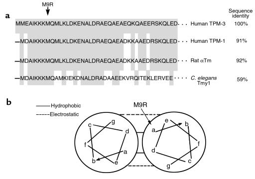
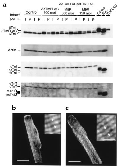
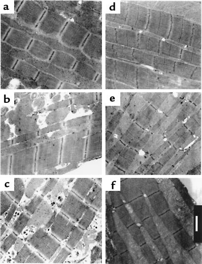
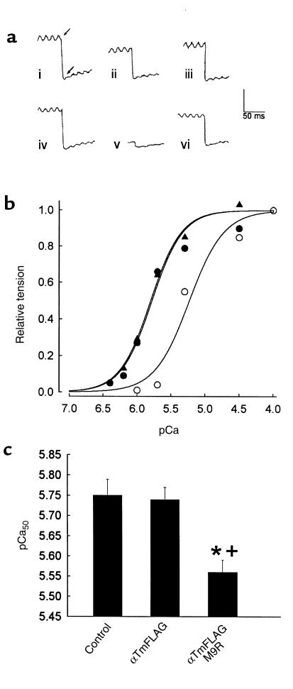
Nemaline myopathy (NM) is a rare autosomal dominant skeletal muscle myopathy characterized by severe muscle weakness and the subsequent appearance of nemaline rods within the muscle fibers. Recently, a missense mutation inTPM3, which encodes the slow skeletal alpha-tropomyosin (alphaTm), was linked to NM in a large kindred with an autosomal-dominant, childhood-onset form of the disease. We used adenoviral gene transfer to fully differentiated rat adult myocytes in vitro to determine the effects of NM mutant human alphaTm expression on striated muscle sarcomeric structure and contractile function. The mutant alphaTm was expressed and incorporated correctly into sarcomeres of adult muscle cells. The primary defect caused by expression of the mutant alphaTm was a decrease in the sensitivity of contraction to activating Ca(2+), which could help explain the hypotonia seen in NM. Interestingly, NM mutant alphaTm expression did not directly result in nemaline rod formation, which suggests that rod formation is secondary to contractile dysfunction and that load-dependent processes are likely involved in nemaline rod formation in vivo.
Figures
Similar articles
-
An alphaTropomyosin mutation alters dimer preference in nemaline myopathy.Ann Neurol. 2005 Jan;57(1):42-9. doi: 10.1002/ana.20305. Ann Neurol. 2005. PMID: 15562513
-
Divergent abnormal muscle relaxation by hypertrophic cardiomyopathy and nemaline myopathy mutant tropomyosins.Physiol Genomics. 2002;9(2):103-11. doi: 10.1152/physiolgenomics.00099.2001. Epub 2002 Mar 26. Physiol Genomics. 2002. PMID: 12006676
-
Alteration of tropomyosin function and folding by a nemaline myopathy-causing mutation.Biophys J. 2000 Dec;79(6):3217-25. doi: 10.1016/S0006-3495(00)76554-4. Biophys J. 2000. PMID: 11106625 Free PMC article.
-
Physiological consequences of tropomyosin mutations associated with cardiac and skeletal myopathies.J Mol Med (Berl). 2000;78(10):543-53. doi: 10.1007/s001090000161. J Mol Med (Berl). 2000. PMID: 11199327 Review.
-
Myopathies associated with β-tropomyosin mutations.Neuromuscul Disord. 2012 Nov;22(11):923-33. doi: 10.1016/j.nmd.2012.05.018. Epub 2012 Jun 29. Neuromuscul Disord. 2012. PMID: 22749895 Review.
Cited by
-
Dilated cardiomyopathy mutations in δ-sarcoglycan exert a dominant-negative effect on cardiac myocyte mechanical stability.Am J Physiol Heart Circ Physiol. 2016 May 1;310(9):H1140-50. doi: 10.1152/ajpheart.00521.2015. Epub 2016 Mar 11. Am J Physiol Heart Circ Physiol. 2016. PMID: 26968544 Free PMC article.
-
Vertebrate tropomyosin: distribution, properties and function.J Muscle Res Cell Motil. 2001;22(1):5-49. doi: 10.1023/a:1010303732441. J Muscle Res Cell Motil. 2001. PMID: 11563548 Review.
-
Congenital myopathy-related mutations in tropomyosin disrupt regulatory function through altered actin affinity and tropomodulin binding.FEBS J. 2019 May;286(10):1877-1893. doi: 10.1111/febs.14787. Epub 2019 Mar 5. FEBS J. 2019. PMID: 30768849 Free PMC article.
-
Defective regulation of contractile function in muscle fibres carrying an E41K beta-tropomyosin mutation.J Physiol. 2008 Jun 15;586(12):2993-3004. doi: 10.1113/jphysiol.2008.153650. Epub 2008 Apr 17. J Physiol. 2008. PMID: 18420702 Free PMC article.
-
Congenital myopathy-causing tropomyosin mutations induce thin filament dysfunction via distinct physiological mechanisms.Hum Mol Genet. 2012 Oct 15;21(20):4473-85. doi: 10.1093/hmg/dds289. Epub 2012 Jul 13. Hum Mol Genet. 2012. PMID: 22798622 Free PMC article.
References
-
- Jockusch BM, et al. Immunofluorescence microscopy of a myopathy. Alpha-actinin is a major constituent of nemaline rods. Exp Cell Res. 1980;127:409–420. - PubMed
-
- Laing NG, et al. A mutation in the α-tropomyosin gene TPM3 associated with autosomal dominant nemaline myopathy. Nat Genet. 1995;9:75–79. - PubMed
-
- Farah CS, Reinach FC. The troponin complex and regulation of muscle contraction. FASEB J. 1995;9:755–767. - PubMed
-
- Tobacman LS. Thin filament-mediated regulation of cardiac contraction. Annu Rev Physiol. 1996;58:447–481. - PubMed
Publication types
MeSH terms
Substances
LinkOut - more resources
Full Text Sources
Molecular Biology Databases
Miscellaneous
