. 1999 Dec;104(11):1621-9.
doi: 10.1172/JCI7515.

Overexpression of monocarboxylate transporter and lactate dehydrogenase alters insulin secretory responses to pyruvate and lactate in beta cells

Affiliations

Overexpression of monocarboxylate transporter and lactate dehydrogenase alters insulin secretory responses to pyruvate and lactate in beta cells

H Ishihara et al. J Clin Invest. 1999 Dec.

Abstract

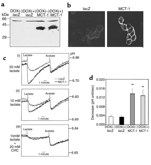
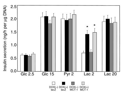
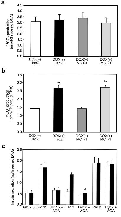
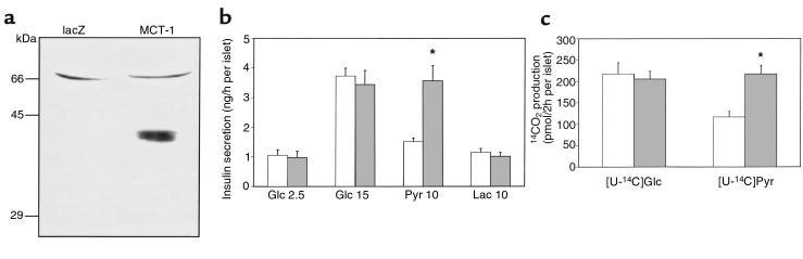
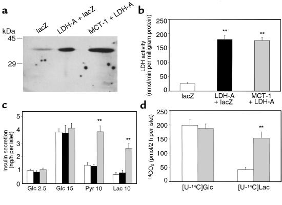
Previous investigations revealed low activities of lactate dehydrogenase (LDH) and plasma membrane monocarboxylate transporters (MCT) in the pancreatic beta cell. In this study the significance of these characteristics was explored by overexpressing type A LDH (LDH-A) and/or type 1 MCT (MCT-1) in the clonal INS-1 beta cells and isolated rat islets. Inducible overexpression of LDH-A resulted in an 87-fold increase in LDH activity in INS-1 cells. Adenovirus-mediated overexpression of MCT-1 increased lactate transport activity 3.7-fold in INS-1 cells. Although overexpression of LDH-A, and/or MCT-1 did not affect glucose-stimulated insulin secretion, LDH-A overexpression resulted in stimulation of insulin secretion even at a low lactate concentration with a concomitant increase in its oxidation in INS-1 cells regardless of MCT-1 co-overexpression. Adenovirus-mediated overexpression of MCT-1 caused an increase in pyruvate oxidation and conferred pyruvate-stimulated insulin release to isolated rat islets. Although lactate did not stimulate insulin secretion from control or MCT-1-overexpressing islets, co-overexpression of LDH-A and MCT-1 evoked lactate-stimulated insulin secretion with a concomitant increase in lactate oxidation in rat islets. These results suggest that low expression of MCT and LDH is requisite to the specificity of glucose in insulin secretion, protecting the organism from undesired hypoglycemic actions of pyruvate and lactate during exercise and other catabolic states.


Figures

Figure 1
Figure 1
Doxycycline-inducible overexpression of LDH-A in INS-1 cells. (a) INS(LDH) cells were infected with either AdCAlacZ or AdCAMCT-1 and cultured in the absence or presence of doxycycline (DOX; 500 ng/mL). Cellular homogenates (100 μg) were dissolved in 11% SDS-PAGE, transferred to nitrocellulose, and immunoblotted with antibody against purified rat liver LDH. (b) LDH activity was determined in cell homogenates using pyruvate as a substrate. Data show the mean ± SEM of 3 independent experiments. **Difference from control cells (noninduced and infected with AdCAlacZ) at P < 0.01.
Figure 2
Figure 2
Overexpression of MCT-1 through a recombinant adenovirus in INS-1 cells. (a) INS(LDH) cells were treated as indicated in Figure 1a. Crude membrane preparations (50 μg) were dissolved in 9% SDS-PAGE and immunoblotted with antibody against COOH-terminal peptide of rat MCT-1. (b) Infected INS(LDH) cells were fixed and immunostained with antibody against MCT-1. (c) Infected INS(LDH) cells were loaded with the pH sensitive dye BCECF and challenged with 20 mM lactate (i) or 10 mM lactate in the absence (ii) or presence (iii) of CHC (20 mM). Traces are representative of more than 4 traces from 2 to 5 experiments. (d) Initial rates of pH decrease were calculated from 4 traces each from 4 different cell preparations. **Difference from control cells at P < 0.01.
Figure 3
Figure 3
Insulin secretion from INS-1 cells overexpressing LDH-A and/or MCT-1. INS(LDH) cells cultured in the presence or absence of doxycycline and infected with either AdCAlacZ or AdCAMCT-1 were incubated in KRBH with the indicated stimuli for 60 minutes. Insulin secreted in KRBH was quantified by radioimmunoassay and normalized by cellular DNA content. Data show the mean ± SEM of 4 or 5 independent experiments. *Difference from control cells (noninduced and infected with AdCAlacZ) at P < 0.05.
Figure 4
Figure 4
Mitochondrial oxidation in INS(LDH) cells overexpressing LDH-A and/or MCT-1. 14CO2 formation during 2-hour incubation with 15 mM [U-14C]glucose (a) or 2 mM [U-14C]lactate (b) were measured. Data show the mean ± SEM of 3 independent experiments. **Difference from control cells (noninduced and infected with AdCAlacZ) at P < 0.01. (c) INS(LDH) cells cultured in the presence (filled bars) or absence (open bars) of doxycycline were incubated in KRBH with the indicated stimuli. AOA (0.25 mM), an inhibitor of the malate-aspartate shuttle, was included where indicated. Statistical significance was obtained (P < 0.01) in lactate-stimulated insulin secretion between AOA-treated and nontreated cells overexpressing LDH-A.
Figure 5
Figure 5
Overexpression of MCT-1 through a recombinant adenovirus and its effects on isolated islets. (a) One hundred twenty islets infected with either AdCAlacZ or AdCAMCT-1 were used for immunoblot analyses. The result is representative of 3 independent experiments. (b) Ten islets infected with recombinant adenoviruses in each tube were incubated in KRBH with indicated stimuli for 60 minutes. Data show the mean ± SEM of 5 independent experiments. Statistical significance between MCT-1–overexpressing islets and control islets was obtained at 10 mM pyruvate (P < 0.05). (c) 14CO2 formation during 2-hour incubation with 15 mM [U-14C]glucose or 10 mM [1-14C]pyruvate was measured. Data show the mean ± SEM of 5 independent experiments. *Difference from control islets at P < 0.05. Open bars, islets infected with AdCAlacZ; shaded bars, islets infected with AdCAMCT-1.
Figure 6
Figure 6
Co-overexpression of MCT-1 and LDH-A through recombinant adenoviruses and its effects on isolated islets. (a) One hundred twenty islets infected with either AdCAlacZ, AdCALDH-A plus AdCAlacZ (AdCAlacZ was combined to adjust total virus amounts), or AdCAMCT-1 plus AdCALDH-A were used for immunoblot analyses. The result is representative of 2 independent experiments. (b) Lysates of 100 islets infected with recombinant adenoviruses were assayed for LDH activity. Data show the mean ± SEM of 3 independent experiments. **Difference from control islets at P < 0.01. (c) Ten islets infected with recombinant adenoviruses in each tube were incubated in KRBH with the indicated stimuli for 60 minutes. Data show the mean ± SEM of 4 independent experiments. Statistical significance between islets overexpressing both MCT-1 and LDH-A and control islets were obtained at 10 mM pyruvate and 10 mM lactate (P < 0.01). (d) 14CO2 formation during 2-hour incubation with 15 mM [U-14C]glucose or 10 mM [U-14C]lactate were measured. Data show the mean ± SEM of 3 independent experiments. **Difference from control islets at P < 0.01. Open bars, islets infected with AdCAlacZ; filled bars, islets infected with AdCALDH-A plus AdCAlacZ; shaded bars, islets infected with AdCAMCT-1 plus AdCALDH-A.

Similar articles

Cited by

References

    1. DeFronzo RA. Lilly Lecture 1987. The triumvirate: β-cell, muscle, liver. A collusion responsible for NIDDM. Diabetes. 1988;37:667–687. - PubMed
    1. Virkarmaki A, Ueki K, Kahn CR. Protein-protein interaction in insulin signaling and the molecular mechanism of insulin resistance. J Clin Invest. 1999;103:931–943. - PMC - PubMed
    1. Prentki M. New insights into pancreatic β-cell metabolic signaling in insulin secretion. Eur J Endocrinol. 1996;134:272–286. - PubMed
    1. Matschinsky FM. Banting Lecture 1995. A lesson in metabolic regulation inspired by the glucokinase glucose sensor paradigm. Diabetes. 1996;45:223–241. - PubMed
    1. MacDonald MJ. Influence of glucose on pyruvate carboxylase expression in pancreatic islets. Arch Biochem Biophys. 1995;319:128–132. - PubMed

Publication types

MeSH terms