

Case Reports
Pseudohemangioma of the vertebra: an unusual radiographic manifestation of primary Ewing's sarcoma
Affiliations
- PMID: 10588101
- PMCID: PMC7657769
Item in Clipboard
Case Reports
Pseudohemangioma of the vertebra: an unusual radiographic manifestation of primary Ewing's sarcoma
AJNR Am J Neuroradiol.
1999 Nov-Dec.
Abstract
Primary Ewing's sarcoma (ES) of the spine is uncommon, exhibiting a variety of appearances on plain-film radiographs and cross-sectional images. We report the unusual CT imaging manifestations of a primary ES with a coarse trabecular pattern that mimicked an aggressive hemangioma of the cervical spine.
Figures
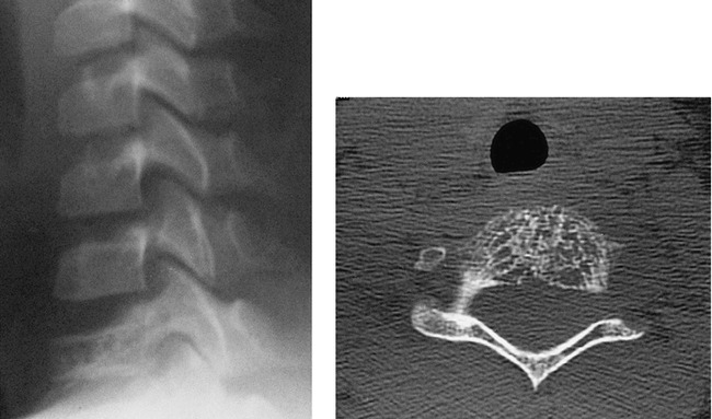
Lateral plain film of lower cervical spine shows compression of C7 vertebra with preservation of C6–7 disk space. fig 2. Axial CT scan without contrast (bone window) shows course trabecular pattern with disruption of cortex of vertebral body. Left neural foramen is slightly widened.
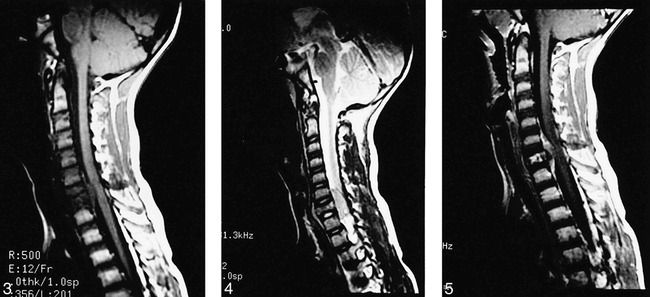
Midline sagittal T1-weighted image of cervical spine shows homogeneous extradural mass compressing spinal cord posteriorly. Mass is low in signal and roughly isointense to gray matter. C7 vertebra is compressed and is decreased in signal with preservation of intervertebral disk spaces. fig 4. Sagittal T2-weighted image of cervical spine slightly to left of midline better shows preservation of intervertebral disk spaces. Extradural mass is fairly homogeneous and intermediate-to-high in signal. Compression of C7 vertebra with increase in anteroposterior diameter is obvious. fig 5. Midline sagittal T1-weighted image with contrast shows heterogeneous enhancement of extradural mass and C7 vertebral body. There is no abnormal enhancement of the spinal cord.
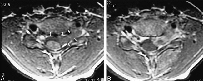
A, T1-weighted axial image through C6–7 intervertebral disk shows extradural mass compressing spinal cord posteriorly. Mass crosses midline and extends out of left neural foramen (arrows). B, T1-weighted axial image with contrast shows heterogeneous enhancement of mass.
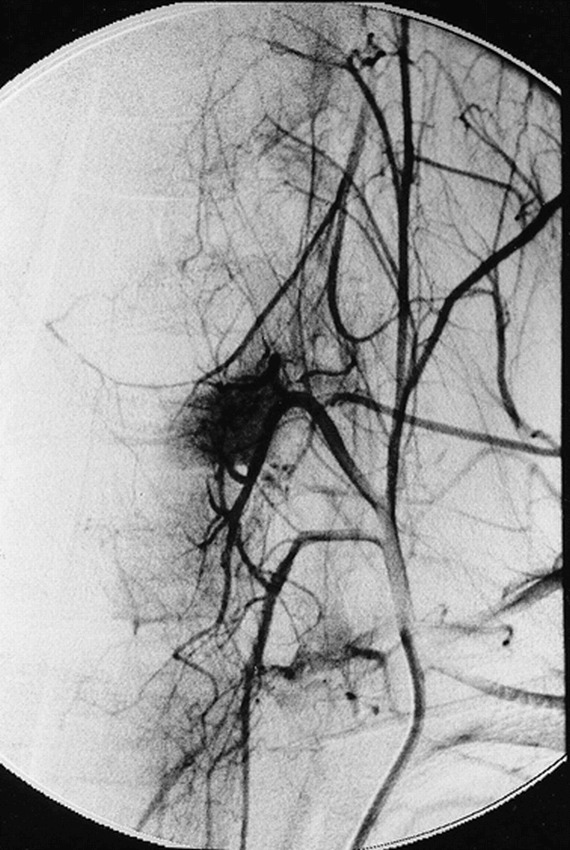
Superselective angiogram of left costocervical trunk. Late arterial-phase projection shows staining in region of epidural mass.
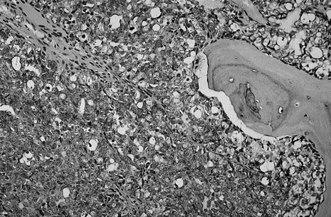
Photomicrograph reveals that tumor is composed of densely packed small, round-to-oval cells (lower left) with high nuclear-to-cytoplasmic ratio (“small blue cell tumor;” several representative cells are highlighted with small arrowheads). Tumor has infiltrated medullary space of vertebrae. Note bone spicule (letter A) on right of figure. Faint thin-walled vascular channels are present (outlined by large arrowheads) (hematoxylin and eosin, original magnification × 200). Cells were immunopositve for CD99 and immunonegative for LCA and PGP 9.5. (not shown).
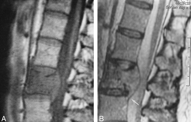
A, Proton density–weighted sagittal image of 53-year-old woman with aggressive VH of L3 vertebra. There is decrease of signal in L3 vertebral body. There is a large, homogeneous, low-signal extraosseous mass within spinal canal compressing thecal sac. There is growth of tumor superiorly and inferiorly along posterior aspect of L3 vertebral body. Note that there is preservation of intervertebral disk spaces. B, T2-weighted sagittal images (from same patient as 9A) shows that signal in extraosseous component increases on T2-weighted image. T2-weighted image better shows how tumor extends beyond inferior disk space (arrow).
Similar articles
-
Intradural, extramedullary spinal Ewing's sarcoma in childhood.J Clin Neurosci. 2003 Jan;10(1):122-5. doi: 10.1016/s0967-5868(02)00279-5. J Clin Neurosci. 2003. PMID: 12464543
-
Primary Ewing's sarcoma of the second cervical vertebra: a rare entity.J Pediatr Orthop B. 2011 Nov;20(6):408-12. doi: 10.1097/bpb.0b013e328345d78a. J Pediatr Orthop B. 2011. PMID: 22272383 No abstract available.
-
Osteochondroma of the cervical spine. MR findings.Clin Imaging. 1995 Oct-Dec;19(4):275-8. doi: 10.1016/0899-7071(94)00063-i. Clin Imaging. 1995. PMID: 8564873
-
Osteochondroma of the cervical spine: case report and review of the literature.Br J Neurosurg. 1994;8(3):359-63. doi: 10.3109/02688699409029627. Br J Neurosurg. 1994. PMID: 7946028 Review.
-
Extraskeletal Ewing's sarcoma: a case report and review of the literature.Spine (Phila Pa 1976). 2000 Aug 1;25(15):1996-9. doi: 10.1097/00007632-200008010-00022. Spine (Phila Pa 1976). 2000. PMID: 10908947 Review.
Cited by
-
Primary Ewing's Sarcoma of the Spine: About a Case.Glob Pediatr Health. 2022 Nov 17;9:2333794X221123874. doi: 10.1177/2333794X221123874. eCollection 2022. Glob Pediatr Health. 2022. PMID: 36420454 Free PMC article.
-
Unusual Presentation of a Primary Ewing's Sarcoma of the Spine with Paraplegia: A Case Report.J Clin Diagn Res. 2015 Mar;9(3):RD01-3. doi: 10.7860/JCDR/2015/11647.5663. Epub 2015 Mar 1. J Clin Diagn Res. 2015. PMID: 25954672 Free PMC article.
-
Vertebroplasty for vertebral hemangioma in children: a report of two cases with 2-year follow-up.Childs Nerv Syst. 2015 Nov;31(11):2179-83. doi: 10.1007/s00381-015-2777-4. Epub 2015 Jun 13. Childs Nerv Syst. 2015. PMID: 26070966
-
Primary Ewing's sarcoma of the vertebral column.Skeletal Radiol. 2004 Sep;33(9):506-13. doi: 10.1007/s00256-004-0810-x. Epub 2004 Jun 30. Skeletal Radiol. 2004. PMID: 15232658
-
Primary Ewing's sarcoma of the spine presenting as acute paraplegia.J Pediatr Neurosci. 2012 Jan;7(1):64-6. doi: 10.4103/1817-1745.97630. J Pediatr Neurosci. 2012. PMID: 22837785 Free PMC article.
References
-
- Amour TE, Hodges SC, Laakman RW, Tamas DE, Other primary extradural tumors. In: MRI of the Spine. Amour TE, Hodges SC, Laakman RW, Tamas DE, eds, New York: Raven Press; 1994:509–516
-
- Grubb MR, Currier BL, Prichard DJ, Ebersold MJ, Primary Ewing's sarcoma of the spine. Spine 1994;19:309-311 - PubMed
-
- Whitehouse GH, Griffiths GJ, Roentgenological aspects of spinal involvement by primary and metastatic Ewing's tumor. J Can Assoc Radiol 1976;27:290-297 - PubMed
-
- Twohig M, Sze G, Spinal tumors. In: Magnetic Resonance Imaging of Children. Cohen MD, Edwards, MK, eds. Philadelphia: BC Decker Inc; 1990:463–498
-
- Fechner RE, Mills SM, Small cell sarcomas. In: Tumors of the Bones and Joints. Fechner RE, Mills SM, eds. Washington, DC: Armed Forces Institute of Pathology; 1993:87–202
Publication types
MeSH terms
LinkOut - more resources
Full Text Sources
Medical