

Syndromes of bilateral symmetrical polymicrogyria
- PMID: 10588102
- PMCID: PMC7657775
Syndromes of bilateral symmetrical polymicrogyria
Abstract
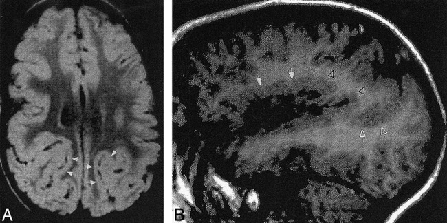
Background and purpose: A number of anatomicoclinical syndromes have been described in which bilateral symmetrical polymicrogyria is the underlying morphologic abnormality. We retrospectively reviewed the clinical, epileptic, and morphologic manifestations of bilateral symmetrical polymicrogyria in 21 patients to determine whether certain areas are at particular risk for these syndromes.
Methods: Clinical records and brain MR studies of 21 patients with bilateral symmetrical polymicrogyria were reviewed to confirm the presence and determine the location of polymicrogyria and to qualitatively correlate location with developmental, neurologic, and epileptic histories. The locations we found were compared with published reports of bilateral symmetrical polymicrogyria to determine whether these locations were random or whether predilections exist for certain areas.
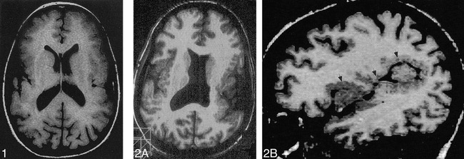
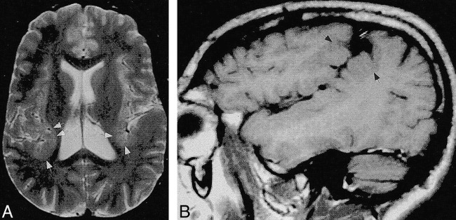
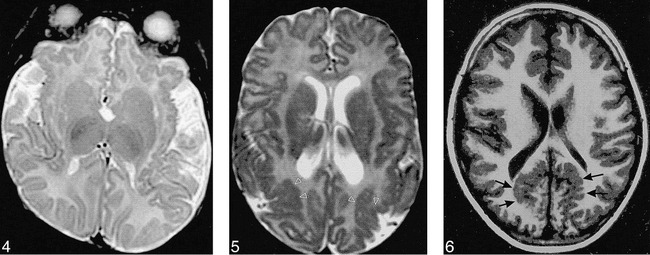
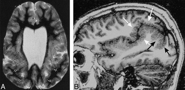
Results: Analysis revealed six patients with bilateral frontal polymicrogyria, nine with bilateral perisylvian polymicrogyria, one with bilateral parietal polymicrogyria, one with bilateral parasagittal parieto-occipital polymicrogyria, two with bilateral frontal polymicrogyria and bilateral perisylvian polymicrogyria, one with bilateral perisylvian and bilateral parasagittal parieto-occipital polymicrogyria, and one with bilateral perisylvian, bilateral parieto-occipital, and bilateral parasagittal parieto-occipital polymicrogyria. Symptom complexes were non-specific, but seemed additive according to the regions of brain involved.
Conclusion: Bilateral symmetrical polymicrogyria has a propensity to develop in specific regions of the cerebral cortex. When the regions are extensive, the areas involved often appear to be simple topological additions of those regions. These locations and the identification of several familial cases raise the possibility that genetic mechanisms influence the development of these malformations in some patients.
Figures
References
-
- Barkovich AJ, Kuzniecky RI, Dobyns WB, Jackson GD, Becker LE, Evrard P, A classification scheme for malformations of cortical development. Neuropediatrics 1996;27:59-63 - PubMed
-
- Guerrini R, Dravet C, Raybaud C, et al. Epilepsy and focal gyral anomalies detected by MRI: electroclinico-morphological correlations and follow-up. Dev Med Child Neurol 1992;34:706-718 - PubMed
-
- Guerrini R, Dravet C, Bureau M, et al. Diffuse and localized dysplasias of cerebral cortex: clinical presentation, outcome, and proposal for a morphologic MRI classification based on a study of 90 patients. In: Guerrini R, Andermann F, Canapicchi R, Roger J, Zifkin BG, Pfanner P, eds. Dysplasias of Cerebral Cortex and Epilepsy. Philadelphia: Lippincott-Raven; 1996: 255–269
-
- Kuzniecky R, Andermann F, Guerrini R, Congenital bilateral perisylvian syndrome; study of 31 patients: the congenital bilateral perisylvian syndrome multicenter collaborative study. Lancet 1993;341:608-612 - PubMed
-
- Kuzniecky R, Morawetz R, Faught E, Black L, Frontal and central lobe focal dysplasia: clinical, EEG, and imaging features. Dev Med Child Neurol 1995;37:159-166 - PubMed
MeSH terms
LinkOut - more resources
Full Text Sources
Medical