Hyperacute ischemic stroke missed by diffusion-weighted imaging
- PMID: 10588111
- PMCID: PMC7657789
Hyperacute ischemic stroke missed by diffusion-weighted imaging
Abstract
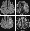
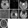
We present two cases of hyperacute ischemic stroke that were initially missed by diffusion-weighted imaging; abnormalities in locations corresponding to focal neurologic deficits were discovered by MR angiography and perfusion-weighted imaging. Within hours, follow-up diffusion-weighted scans revealed partial conversion of the hypoperfused regions to complete stroke. These cases illustrate the potential for a nonresolving stroke-in-evolution to go undetected by diffusion-weighted imaging at hyperacute timepoints.
Figures


Comment in
-
The need for objective assessment of the new imaging techniques and understanding the expanding roles of stroke imaging.AJNR Am J Neuroradiol. 1999 Nov-Dec;20(10):1779-84. AJNR Am J Neuroradiol. 1999. PMID: 10588097 Free PMC article. Review. No abstract available.
Similar articles
-
Diffusion-weighted magnetic resonance imaging in internal carotid artery dissection.Arch Neurol. 2004 Apr;61(4):510-2. doi: 10.1001/archneur.61.4.510. Arch Neurol. 2004. PMID: 15096398
-
Diffusion-negative stroke: a report of two cases.AJNR Am J Neuroradiol. 1999 Nov-Dec;20(10):1876-80. AJNR Am J Neuroradiol. 1999. PMID: 10588112 Free PMC article.
-
Various patterns of perfusion-weighted MR imaging and MR angiographic findings in hyperacute ischemic stroke.AJNR Am J Neuroradiol. 1999 Apr;20(4):613-20. AJNR Am J Neuroradiol. 1999. PMID: 10319971 Free PMC article.
-
[Diffusion-weighted imaging in acute stroke].Nervenarzt. 1998 Aug;69(8):683-93. doi: 10.1007/s001150050329. Nervenarzt. 1998. PMID: 9757420 Review. German.
-
[The clinical application of diffusion weighted magnetic resonance imaging to acute cerebrovascular disorders].No To Shinkei. 1998 Sep;50(9):787-95. No To Shinkei. 1998. PMID: 9789301 Review. Japanese.
Cited by
-
Use of magnetic resonance imaging to predict outcome after stroke: a review of experimental and clinical evidence.J Cereb Blood Flow Metab. 2010 Apr;30(4):703-17. doi: 10.1038/jcbfm.2010.5. Epub 2010 Jan 20. J Cereb Blood Flow Metab. 2010. PMID: 20087362 Free PMC article. Review.
-
Impact of additional resection on new ischemic lesions and their clinical relevance after intraoperative 3 Tesla MRI in neuro-oncological surgery.Neurosurg Rev. 2021 Aug;44(4):2219-2227. doi: 10.1007/s10143-020-01399-9. Epub 2020 Sep 30. Neurosurg Rev. 2021. PMID: 32996078 Free PMC article.
-
Arterial hyperintensity on fast fluid-attenuated inversion recovery images: a subtle finding for hyperacute stroke undetected by diffusion-weighted MR imaging.AJNR Am J Neuroradiol. 2001 Apr;22(4):632-6. AJNR Am J Neuroradiol. 2001. PMID: 11290469 Free PMC article.
-
The role of functional MR imaging in patients with ischemia in the visual cortex.AJNR Am J Neuroradiol. 2001 Jun-Jul;22(6):1043-9. AJNR Am J Neuroradiol. 2001. PMID: 11415895 Free PMC article.
-
Transparent color-coded three-dimensional diffusion-weighted magnetic resonance imaging for the numerical analysis of reversible and irreversible hyperacute ischemic stroke--case report.Neuroradiology. 2005 Apr;47(4):245-50. doi: 10.1007/s00234-004-1315-y. Epub 2005 Apr 2. Neuroradiology. 2005. PMID: 15806432
References
-
- Pierpaoli C, Righini A, Linfante I, et al. Histopathologic correlates of abnormal water diffusion in cerebral ischemia: diffusion-weighted MR imaging and light and electron microscopic study. Radiology 1993;189:439-448 - PubMed
-
- Pierpaoli C, Alger JR, Righini A, et al. High temporal resolution diffusion MRI of global cerebral ischemia and reperfusion. J Cereb Blood Flow Metab 1996;16:892-905 - PubMed
-
- Gonzalez G, Schaefer P, Buonanno F, et al. Clinical sensitivity and specificity of diffusion weighted MRI in hyperacute stroke. American Heart Association 22nd International Joint Conference on Stroke and Cerebral Circulation; February 6–8, 1997; Anaheim, Calif
-
- Barber PA, Darby DG, Yang Q, et al. Echoplanar magnetic resonance imaging (EPI) perfusion and diffusion abnormalities correlate with outcome in acute stroke. American Heart Association 23rd International Joint Conference on Stroke and Cerebral Circulation; February 5–7, 1998; Orlando, Fl
Publication types
MeSH terms
LinkOut - more resources
Full Text Sources
Medical
