

Central processing of rectal pain: a functional MR imaging study
- PMID: 10588119
- PMCID: PMC7657788
Central processing of rectal pain: a functional MR imaging study
Abstract
Background and purpose: Although the central processing of somatic pain has been dealt with in numerous brain imaging studies, the neural correlates of visceral pain have received much more limited attention. Our goal was to assess the feasibility of detecting brain activation patterns induced by rectal pain by means of functional MR imaging. We hypothesized that the cerebral processing of rectal pain would exhibit strong similarities with the central processing of somatic pain.
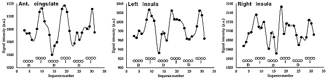
Methods: Functional MR imaging data were obtained from eight healthy subjects. A block paradigm was applied. Rectal pain was induced by inflating a latex balloon catheter that had been inserted into the rectum. Functional responses were established by means of cross-correlation analysis.
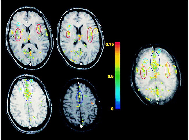
Results: Activation was detected within the anterior cingulate gyrus, the prefrontal cortex, the insular cortex, the sensory-motor cortex, the inferior parietal lobule, the posterior cingulate gyrus, and the visual cortex.
Conclusion: Functional MR imaging of visceral pain is feasible in healthy subjects. The activation patterns observed in this study support the hypothesis that the cerebral processing of visceral pain involves multiple components, similar to the central processing of somatic pain. Our results constitute a first step toward the identification of possible aberrations in the activation patterns of patients suffering from visceral hypersensitivity.
Figures
References
-
- Jones AKP, Friston KJ, Frackowiak RSJ, Cerebral localisation of responses to pain in man using positron emission tomography. Science 1992;255:215-216 - PubMed
-
- Talbot JD, Marrett S, Evans AC, Meyer E, Bushnell MC, Duncan GH, Multiple representations of pain in human cerebral cortex. Science 1991;251:1355-1358 - PubMed
-
- Rainville P, Duncan GH, Price DD, Carrier B, Bushnell C, Pain affect encoded in human anterior cingulate but not somatosensory cortex. . Science 1997;277:968-971 - PubMed
-
- Casey KL, Minoshima S, Morrow TJ, Koeppe RA, Comparison of human cerebral activation patterns during cutaneous warmth, heatpain and deep cold pain. . J Neurophysiol 1996;76:571-581 - PubMed
-
- Vogt BA, Derbyshire S, Jones AKP, Pain processing in four regions of human cingulate cortex localized with co-registered PET and MR imaging. . Eur J Neurosci 1996;8:1461-1473 - PubMed
Publication types
MeSH terms
LinkOut - more resources
Full Text Sources
Medical