

Evolution of multiple sclerosis lesions on serial contrast-enhanced T1-weighted and magnetization-transfer MR images
- PMID: 10588122
- PMCID: PMC7657782
Evolution of multiple sclerosis lesions on serial contrast-enhanced T1-weighted and magnetization-transfer MR images
Abstract
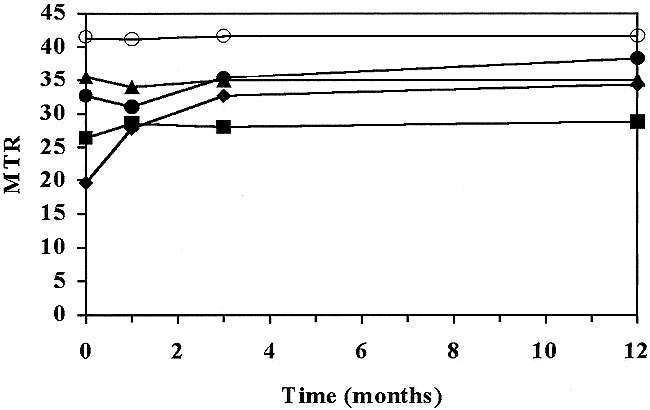
Background and purpose: Magnetization-transfer imaging is a technique that could provide indirect evidence of the characteristics of multiple sclerosis (MS) lesions. The purpose of this work was to study the evolution of MS lesions on T1-weighted MR images over time and to investigate changes in magnetization-transfer ratio (MTR) values of MS lesions with different initial appearances on contrast-enhanced T1-weighted images.
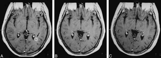
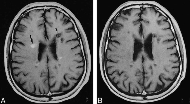
Methods: Eleven patients with relapsing-remitting MS were studied with MR imaging. The MTRs were calculated for 47 lesions that had been classified according to their appearance on contrast-enhanced T1-weighted images. Each patient was examined at four time points over a 1-year period. The MTR changes observed in the selected lesions were compared with their initial T1-weighted appearance.
Results: The lowest MTR values were initially found in hypointense nonenhancing lesions and in ring-enhancing lesions, with both types showing a hypointense center. Changes in MTR values were more dynamic and reversible in ring-enhancing than in hypointense nonenhancing plaques. Nodular-enhancing lesions had slightly lower initial MTRs than did isointense non-enhancing lesions.
Conclusion: The absence or presence of contrast uptake may indicate a different pathologic basis for hypointense MS lesions on T1-weighted MR images. These differences should be kept in mind when considering T1 lesion load as a surrogate marker of disability in MS.
Figures

Similar articles
-
Patterns of lesion development in multiple sclerosis: longitudinal observations with T1-weighted spin-echo and magnetization transfer MR.AJNR Am J Neuroradiol. 1998 Apr;19(4):675-83. AJNR Am J Neuroradiol. 1998. PMID: 9576653 Free PMC article.
-
Coefficient D(av) is more sensitive than fractional anisotropy in monitoring progression of irreversible tissue damage in focal nonactive multiple sclerosis lesions.AJNR Am J Neuroradiol. 2003 Apr;24(4):663-70. AJNR Am J Neuroradiol. 2003. PMID: 12695200 Free PMC article.
-
Magnetization transfer effects in MR-detected multiple sclerosis lesions: comparison with gadolinium-enhanced spin-echo images and nonenhanced T1-weighted images.AJNR Am J Neuroradiol. 1995 Jan;16(1):69-77. AJNR Am J Neuroradiol. 1995. PMID: 7900604 Free PMC article.
-
Characterization of multiple sclerosis plaques with T1-weighted MR and quantitative magnetization transfer.AJNR Am J Neuroradiol. 1995 Aug;16(7):1473-9. AJNR Am J Neuroradiol. 1995. PMID: 7484636 Free PMC article.
-
The role of magnetic resonance techniques in understanding and managing multiple sclerosis.Brain. 1998 Jan;121 ( Pt 1):3-24. doi: 10.1093/brain/121.1.3. Brain. 1998. PMID: 9549485 Review.
Cited by
-
Advanced magnetic resonance imaging techniques to better understand multiple sclerosis.Biophys Rev. 2010 May;2(2):83-90. doi: 10.1007/s12551-010-0031-6. Epub 2010 Apr 2. Biophys Rev. 2010. PMID: 28510010 Free PMC article.
-
Quantitative magnetization transfer imaging in relapsing-remitting multiple sclerosis: a systematic review and meta-analysis.Brain Commun. 2022 Apr 4;4(2):fcac088. doi: 10.1093/braincomms/fcac088. eCollection 2022. Brain Commun. 2022. PMID: 35652121 Free PMC article. Review.
-
MRI in multiple sclerosis: what's inside the toolbox?Neurotherapeutics. 2007 Oct;4(4):602-17. doi: 10.1016/j.nurt.2007.08.001. Neurotherapeutics. 2007. PMID: 17920541 Free PMC article. Review.
-
Use of Magnetic Resonance Imaging as Well as Clinical Disease Activity in the Clinical Classification of Multiple Sclerosis and Assessment of Its Course: A Report from an International CMSC Consensus Conference, March 5-7, 2010.Int J MS Care. 2012 Fall;14(3):105-14. doi: 10.7224/1537-2073-14.3.105. Int J MS Care. 2012. PMID: 24453741 Free PMC article.
-
Longitudinal analysis of new multiple sclerosis lesions with magnetization transfer and diffusion tensor imaging.Eur Radiol. 2024 Mar;34(3):1680-1691. doi: 10.1007/s00330-023-10173-6. Epub 2023 Sep 2. Eur Radiol. 2024. PMID: 37658894 Free PMC article.
References
-
- Whitaker JN, McFarland HF, Rudge P, Reingold SC, Outcomes assessment in multiple sclerosis clinical trials: a critical analysis. Multiple Sclerosis 1995;1:37-47 - PubMed
-
- Miller DH, Albert PS, Barkhof F, et al. Guidelines for the use of magnetic resonance techniques in monitoring the treatment of multiple sclerosis. Ann Neurol 1996;39:6-16 - PubMed
-
- Miller DH, Grossman RI, Reingold SC, McFarland HF, The role of magnetic resonance techniques in understanding and managing multiple sclerosis. Brain 1998;121:3-24 - PubMed
-
- Runge VM, Price AC, Kirshner HS, et al. MR imaging of MS: a study of pulse-techniques efficiency. AJR Am J Roentgenol 1984;143:1015-1026 - PubMed
-
- Ormerod IEC, Miller DH, McDonald WI, et al. The role of NMR imaging in the assessment of multiple sclerosis and isolated neurological lesions: a quantitative study. Brain 1987;110:1579-1616 - PubMed
Publication types
MeSH terms
Substances
LinkOut - more resources
Full Text Sources
Medical