

Case Reports
Kimura's disease with bilateral auricular masses
Affiliations
- PMID: 10588129
- PMCID: PMC7657797
Item in Clipboard
Case Reports
Kimura's disease with bilateral auricular masses
AJNR Am J Neuroradiol.
1999 Nov-Dec.
Abstract
We report an unusual case of Kimura's disease. An 81-year-old Japanese woman was shown to have bilateral auricular masses that had begun to enlarge 6 years before. On CT scans, slightly high-density masses with faint contrast enhancement were seen. The masses were heterogeneous and hypointense on T1-weighted MR images, were slightly hyperintense on T2-weighted MR images, and showed heterogeneous enhancement after the administration of contrast material. Kimura's disease should be included in the differential diagnosis of bilateral auricular tumors.
Figures
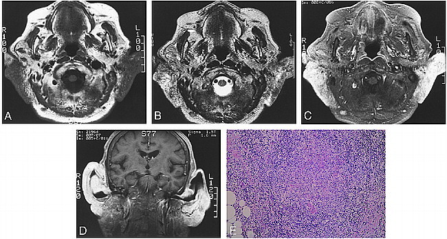
Images from the case of an 81-year-old Japanese woman who was admitted to our hospital with complaints of almost synchronous bilateral auricular masses that had begun to enlarge 6 years before admission and were accompanied by itchiness. A, T1-weighted MR image (440/19/2 [TR/TE/excitations]) shows irregular masses in the bilateral auricles, parotid glands, and retroauricular spaces, which are heterogeneous and hypointense compared with the muscles. B, T2-weighted MR image (4000/108/2) shows that the masses are hyperintense. C, After the administration of contrast material, a T1-weighted axial view image (540/16/1.5) shows heterogeneously enhancing masses involving both auricles. Enlarged bilateral cervical lymph nodes were also noted and showed homogeneous enhancement. D, Contrast-enhanced T1-weighted coronal view image (400/11/2) shows heterogeneously enhancing masses involving both auricles. E, Microscopically, marked infiltration of eosinophils and lymphocytes with germinal centers and proliferation of many blood vessels and thick fibrotic tissues are seen, compatible with Kimura's disease (hematoxylin and eosin stain; original magnification, ×40).
References
-
- Kase Y, Ikeda T, Yamane M, et al. Kimura's disease. Report of 4 cases with a review of 130 reported cases. Otolaryngol Head Neck Surg 1990;63:413-418
-
- Li T-J, Chen X-M, Wang S-Z, et al. Kimura's disease. Oral Surg Oral Med Oral Pathol Oral Radiol Endod 1996;82:549-555 - PubMed
-
- Hirokawa Y, Ikeda K, Ha-Kawa SK, et al. A case of eosinophilic lymphoid granuloma with fibrosis. Jpn J Clin Radiol 1987;42:357-360
-
- Weiss LM, Chan WC, Schnitzer B, Anderson's Pathology.. 10th ed. St. Louis: Mosby; 1996:1137
Publication types
MeSH terms
LinkOut - more resources
Full Text Sources
Medical