. 1999 Dec;10(12):4059-73.
doi: 10.1091/mbc.10.12.4059.

Degradation of a short-lived glycoprotein from the lumen of the endoplasmic reticulum: the role of N-linked glycans and the unfolded protein response

Affiliations
Free PMC article

Degradation of a short-lived glycoprotein from the lumen of the endoplasmic reticulum: the role of N-linked glycans and the unfolded protein response

M de Virgilio et al. Mol Biol Cell. 1999 Dec.
Free PMC article

Abstract

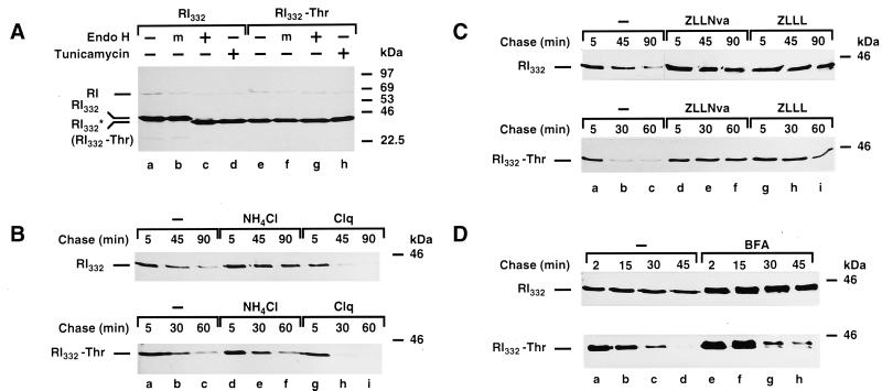
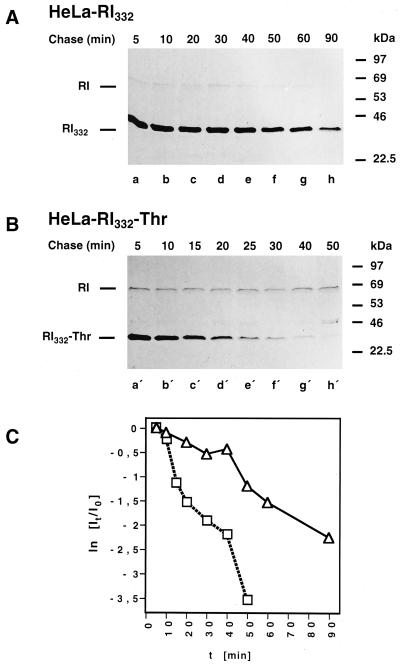
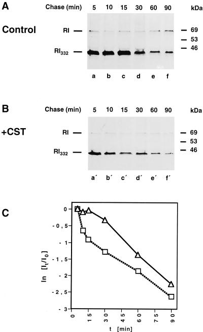
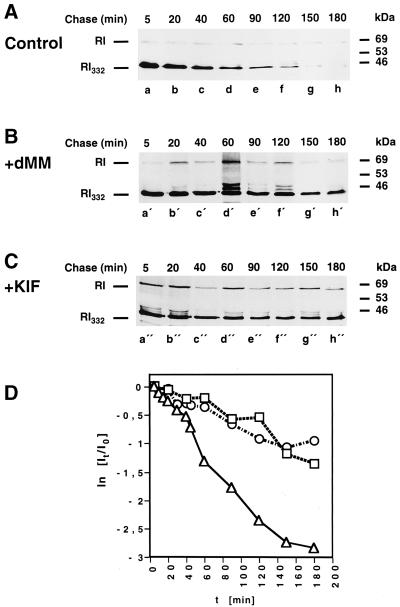
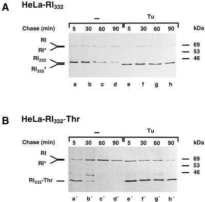
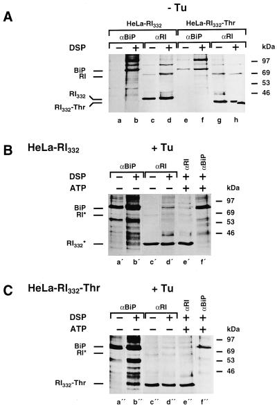
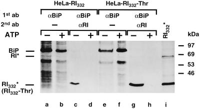
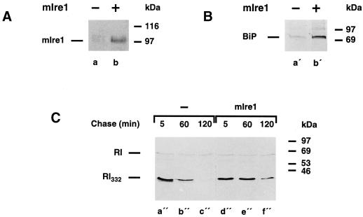
We are studying endoplasmic reticulum-associated degradation (ERAD) with the use of a truncated variant of the type I ER transmembrane glycoprotein ribophorin I (RI). The mutant protein, RI(332), containing only the N-terminal 332 amino acids of the luminal domain of RI, has been shown to interact with calnexin and to be a substrate for the ubiquitin-proteasome pathway. When RI(332) was expressed in HeLa cells, it was degraded with biphasic kinetics; an initial, slow phase of approximately 45 min was followed by a second phase of threefold accelerated degradation. On the other hand, the kinetics of degradation of a form of RI(332) in which the single used N-glycosylation consensus site had been removed (RI(332)-Thr) was monophasic and rapid, implying a role of the N-linked glycan in the first proteolytic phase. RI(332) degradation was enhanced when the binding of glycoproteins to calnexin was prevented. Moreover, the truncated glycoprotein interacted with calnexin preferentially during the first proteolytic phase, which strongly suggests that binding of RI(332) to the lectin-like protein may result in the slow, initial phase of degradation. Additionally, mannose trimming appears to be required for efficient proteolysis of RI(332). After treatment of cells with the inhibitor of N-glycosylation, tunicamycin, destruction of the truncated RI variants was severely inhibited; likewise, in cells preincubated with the calcium ionophore A23187, both RI(332) and RI(332)-Thr were stabilized, despite the presence or absence of the N-linked glycan. On the other hand, both drugs are known to trigger the unfolded protein response (UPR), resulting in the induction of BiP and other ER-resident proteins. Indeed, only in drug-treated cells could an interaction between BiP and RI(332) and RI(332)-Thr be detected. Induction of BiP was also evident after overexpression of murine Ire1, an ER transmembrane kinase known to play a central role in the UPR pathway; at the same time, stabilization of RI(332) was observed. Together, these results suggest that binding of the substrate proteins to UPR-induced chaperones affects their half lives.


Figures

Figure 1
Figure 1
Nonlysosomal and proteasome-dependent degradation of the glycosylated and nonglycosylated ribophorin I variants, RI332 and RI332-Thr. (A) HeLa-RI332 and HeLa-RI332-Thr cells, plated in 35-mm dishes to a density of 5–8 × 10⁵ cells per dish, were left untreated (lanes a–c and e–g) or treated with tunicamycin (5 μg/ml) for 1 h (lanes d and h). The cells were then incubated in serum- and methionine-free medium for 30 min and metabolically labeled with the same medium containing [35S]methionine (250 μCi/ml) for 30 min in the continued absence or presence of the drug. Cells were lysed with SDS-containing buffer and processed for immunoprecipitation with a polyclonal rabbit anti-ribophorin I antibody. The immunoprecipitates were left untreated (lanes a, d, e, and h), mock treated (m; lanes b and f), or digested with endo H (lanes c and g) overnight at 37°C. Finally, all samples were analyzed by SDS-PAGE (10% gels) followed by fluorography. (B and C) HeLa-RI332 and HeLa-RI332-Thr cells were left untreated (B and C, lanes a–c) or preincubated with NH4Cl (50 mM; B, lanes d–f), chloroquine (Clq, 0.1 mM; B, lanes g–i), ZLLNva (40 μM; C, lanes d–f), or ZLLL (50 μM; C, lanes g–i). (D) Cells were incubated in serum- and methionine-free medium in the absence (lanes a–d) or presence of BFA (5 μg/ml; lanes e–h) for 30 min. (B–D) Pulse labeling was carried out in the same medium containing [35S]methionine (250 μCi/ml) for 10 min (B and C) or 5 min (D), followed by chase incubations in complete medium supplemented with unlabeled l-methionine (5 mM) in the continued absence or presence of the drugs for the times indicated. Cell lysis, immunoprecipitations, and sample analysis were performed as described for panel A. Note that because of the lower incorporation of [35S]methionine in the presence of chloroquine, the truncated ribophorins are less apparent already at 45 and 30 min of chase. RI indicates the position of the native ribophorin I, and RI332* indicates the position of nonglycosylated RI332, observed in the presence of tunicamycin.
Figure 2
Figure 2
Kinetics of degradation of RI332 and RI332-Thr expressed in HeLa cells. HeLa-RI332 (A) and HeLa-RI332-Thr (B) cells were pulse labeled for 10 min and chased for up to 90 or 50 min, respectively. The cells were lysed, and anti-ribophorin I immunoprecipitations were performed. All samples were analyzed by SDS-PAGE followed by fluorography. (C) Quantitations of the bands corresponding to radiolabeled full-length (RI) and truncated ribophorin I variants were obtained by scanning densitometry. Semilog plots showing the degradation kinetics of RI332 (▵) and RI332-Thr (□) were established as described in MATERIALS AND METHODS. Data from three independent experiments are shown.
Figure 3
Figure 3
The initial, slow phase of RI332 degradation is omitted in the presence of castanospermine. HeLa-RI332 cells were kept untreated (A) or preincubated with castanospermine (CST; 1 mM) (B). All cell cultures were pulse labeled for 10 min and chased for up to 90 min in complete medium in the continued absence or presence of the drug. Cells were lysed, and RI332 and ribophorin I were immunoprecipitated and analyzed by SDS-PAGE and fluorography. (C) Quantitations of the bands corresponding to radiolabeled ribophorin I (RI) and RI332 to establish the degradation kinetics of RI332 in the absence (▵) or presence (□) of castanospermine were performed as described in the legend of Figure 2C and in MATERIALS AND METHODS. Data from six independent experiments are shown.
Figure 4
Figure 4
RI332 interacts with calnexin only in the absence of castanospermine. HeLa-RI332 (lanes a–f) and HeLa-RI332-Thr (lanes g and h) cells were preincubated, pulse labeled for 10 min, and chased for the times indicated in the absence (lanes a, c, e, and g) or in the presence (lanes b, d, f, and h) of castanospermine (CST; 1 mM). Cells were lysed in buffer containing CHAPS (2%), and anti-calnexin immunoprecipitations were performed under nonstringent conditions in the presence of 1% of the same detergent. The second steps of the sequential immunoprecipitations were carried out under stringent conditions in the presence of SDS (0.6%) and Triton X-100 (1%). Only ribophorin I (RI) and RI332 or RI332-Thr reprecipitated under stringent conditions from anti-calnexin immunoprecipitations obtained under nonstringent conditions are shown for each chase time point. As a control, RI332 and RI332-Thr were immunoprecipitated under stringent conditions from cell lysates of HeLa-RI332 and HeLa-RI332-Thr cells metabolically labeled for 10 min (lanes i and j, respectively). All samples were analyzed by SDS-PAGE and fluorography. Exposure times: lanes a and b, 7 d; lanes c, d, and f–h, 10 d; lane e, 30 d; lanes i and j, 1 d.
Figure 5
Figure 5
RI332 is stabilized in kifunensine- or deoxymannojirimycin-treated HeLa cells. HeLa-RI332 cells were left untreated (A) or preincubated with deoxymannojirimycin (dMM; 2 mM) (B) or kifunensine (KIF; 2 μg/ml) (C). The cells were pulse labeled for 10 min and chased for up to 3 h in the continued absence or presence of the drugs. Anti-ribophorin I immunoprecipitations performed on cell lysates were analyzed by SDS-PAGE and fluorography. (D) The kinetics of RI332 degradation in the absence (▵) or in the presence of dMM (□) or KIF (○) were established as described in the legend of Figure 2 and in MATERIALS AND METHODS. Data from three independent experiments are shown.
Figure 6
Figure 6
The truncated ribophorin I variants are stabilized in tunicamycin-treated HeLa cells. HeLa-RI332 (A) and HeLa-RI332-Thr (B) cells were left untreated (lanes a–d and a′–d′) or preincubated with tunicamycin (Tu, 5 μg/ml; lanes e–h and e′–h′). The cells were pulse labeled for 10 min and chased for up to 90 min in the continued absence or presence of the drug. Anti-ribophorin I immunoprecipitations performed on cell lysates were analyzed by SDS-PAGE and fluorography. RI* and RI332* indicate the positions of nonglycosylated endogenous ribophorin I and RI332, respectively, observed in the presence of tunicamycin.
Figure 7
Figure 7
The synthesis of BiP is highly induced by tunicamycin treatment in HeLa cells. (A) HeLa-RI332 and HeLa-RI332-Thr cells were left untreated (lanes a and e) or preincubated with tunicamycin (Tu; 5 μg/ml) for 1 h (lanes b and f), 2 h (lanes c and g), or 4 h (lanes d and h). Cells were pulse labeled in the continued absence or presence of the drug for 30 min. Cell lysis and immunoprecipitations with the monoclonal mouse anti-BiP antibody were performed as described in MATERIALS AND METHODS. Samples were analyzed by SDS-PAGE and fluorography. (B) HeLa-RI332 and HeLa- RI332-Thr cells, grown in 10-cm dishes, were left untreated (lanes a′ and e′) or preincubated with tunicamycin (5 μg/ml) for the times indicated (lanes b′–d′ and f′–h′). Cell extracts were prepared in Triton X-100–containing buffer, and the pellets were dissolved in an SDS-containing buffer. Cell extracts (20 μg of total protein, corresponding to approximately one-tenth of each extract; lanes a′–d′) and corresponding amounts of the dissolved pellets (lanes e′–h′) were subjected to SDS-PAGE. After transfer of the proteins to nitrocellulose, immunodetection was performed with the use of the anti-BiP antibody and the ECL kit. (C) HeLa-RI332 cells, grown in 6-cm dishes, were preincubated and pulse labeled for 30 min in the absence (lanes a" and c") or presence of tunicamycin (5 μg/ml; lanes b" and d"). Anti-BiP immunoprecipitates obtained from cell lysates were subjected to SDS-PAGE, and the proteins were transferred to a nitrocellulose membrane. The membrane was probed by Western blot analysis with the use of anti-BiP antibodies (lanes c" and d"), and after decay of the signal generated by the ECL reaction, the immunoprecipitates were analyzed by autoradiography with the use of BioMax MR x-ray film (lanes a" and b").
Figure 8
Figure 8
RI332 and RI332-Thr are readily cross-linked to BiP in tunicamycin-treated cells. HeLa-RI332 (A, lanes a–d, and B) and HeLa-RI332-Thr (A, lanes e–h, and C) cells were pretreated and pulse labeled for 30 min in the absence (A) or in the presence (B and C) of tunicamycin (Tu; 5 μg/ml). Cells were lysed in the presence of digitonin (0.2%) and incubated in the absence or presence of DSP (100 μg/ml) as indicated. As a control, a cross-linking experiment was carried out on a cell lysate preincubated with ATP (5 mM; B, lanes e′ and f′; C, lanes e" and f"). Ribophorin I (RI) and RI332 or RI332-Thr were immunoprecipitated from the cell lysates in the presence of SDS and Triton X-100 (A, lanes c, d, g, and h; B, lanes c′–e′; C, lanes c"–e"). Anti-BiP immunoprecipitations were performed under the same conditions (A, lanes a, b, e, and f; B, lanes a′, b′, and f′; C, lanes a", b", and f"). All samples were analyzed by SDS-PAGE followed by fluorography. Exposure times: A, 7 d; B, lanes a′, b′, and f′, 2 d; lanes c′–e′, 7 d; C, lanes a", b", and f", 1 d; lanes c"–e", 7 d.
Figure 9
Figure 9
RI332 and RI332-Thr are coimmunoprecipitated with BiP in tunicamycin-treated HeLa cells. HeLa-RI332 and HeLa-RI332-Thr cells were preincubated with tunicamycin (5 μg/ml) and pulse labeled for 30 min in the presence of the drug. Cell lysis, a subsequent 30-min incubation in the absence (lanes a, c, e, and g) or presence (lanes b, d, f, and h) of ATP (5 mM), and anti-BiP immunoprecipitations were performed as described in MATERIALS AND METHODS. For some samples, the anti-BiP immunoprecipitates were eluted from the protein A–Sepharose beads with a buffer containing SDS (2%) and used for reprecipitations under stringent conditions (lanes c, d, g, and h). One sample was subjected to anti-ribophorin I immunoprecipitation without prior anti-BiP precipitation (lane i). All samples were analyzed by SDS-PAGE and fluorography. Exposure times: lanes a, b, e, f, and i, 20 h; lanes c, d, g, and h, 14 d.
Figure 10
Figure 10
RI332 and RI332-Thr are stabilized after treatment of cells with A23187 and are coimmunoprecipitated with BiP only in the presence of the drug. (A) HeLa-RI332 and HeLa-RI332-Thr cells were left untreated (lanes a–d) or preincubated with A23187 (5 μM) (lanes e–h). The cells were pulse labeled for 10 min and chased for up to 2 h in the continued absence or presence of the drug. Anti-ribophorin I immunoprecipitations performed on cell lysates were analyzed by SDS-PAGE and fluorography. (B) HeLa-RI332 (lanes a′–c′) and HeLa-RI332-Thr (lanes d′–f′) cells were left untreated (lanes a′ and d′) or preincubated with A23187 for 30 min (lanes b′ and e′) or 90 min (lanes c′ and f′). Cells were pulse labeled in the continued absence or presence of the drug for 30 min. Cell lysis and immunoprecipitations with the monoclonal anti-BiP antibody were performed as described in MATERIALS AND METHODS. (C) HeLa-RI332 (lanes a"–c") and HeLa-RI332-Thr (lanes d"–f") cells were preincubated in the absence (lanes a" and d") or in the presence of A23187 (lanes b", c", e", and f") and pulse labeled for 30 min in the continuous absence or presence of the drug. Cell lysis, a 30-min incubation in the absence (lanes a", b", d", and e") or presence (lanes c" and f") of ATP (5 mM), and anti-BiP immunoprecipitations were performed as described in MATERIALS AND METHODS. The anti-BiP immunoprecipitates were eluted from the protein A–Sepharose beads with a buffer containing SDS (2%) and used for a second round of anti-ribophorin I immunoprecipitations under stringent conditions. All samples were analyzed by SDS-PAGE and fluorography. Exposure times: A and B, 2 d; C, 42 d.
Figure 11
Figure 11
mIre1 overexpression induces BiP and prolongs the half life of RI332. (A) Triton X-100 extracts prepared from HeLa-RI332 cells (lane a) and from HeLa-RI332 cells transiently overexpressing mIre1 (lane b) were used to detect the c-myc–tagged version of mIre1 by Western blot analysis. (B) HeLa-RI332 cells (lane a′) and HeLa-RI332 cells transiently overexpressing mIre1 (lane b′) were pulse labeled for 30 min. Anti-BiP immunoprecipitations were performed on lysates prepared from these cells and analyzed by SDS-PAGE followed by fluorography. (C) HeLa-RI332 cells (lanes a"–c") and HeLa-RI332 cells transiently overexpressing mIre1 (lanes d"–f") were pulse labeled for 10 min and chased for up to 120 min. Anti-ribophorin I immunoprecipitations were performed on cell lysates and analyzed by SDS-PAGE and fluorography.

Similar articles

Cited by

References

    1. Amara JF, Lederkremer G, Lodish HF. Intracellular degradation of unassembled asialoglycoprotein receptor subunits: a preGolgi, nonlysosomal endoproteolytic cleavage. J Cell Biol. 1989;109:3315–3324. - PMC - PubMed
    1. Brodsky JL, McCracken AA. ER-associated and proteasome-mediated protein degradation: how two topologically restricted events come together. Trends Cell Biol. 1997;7:151–156. - PubMed
    1. Chang SC, Wooden SK, Nakaki T, Kim YK, Lin AY, Kung L, Attenello JW, Lee AS. Rat gene encoding the 78-kDa glucose-regulated protein GRP78: its regulatory sequences and the effect of protein glycosylation on its expression. Proc Natl Acad Sci USA. 1987;84:680–684. - PMC - PubMed
    1. Chapman R, Sidrauski C, Walter P. Intracellular signaling from the endoplasmic reticulum to the nucleus. Annu Rev Cell Dev Biol. 1998;14:459–485. - PubMed
    1. Chen Y, Caherec F, Chuck SL. Calnexin and other factors that alter translocation affect the rapid binding of ubiquitin to apoB in the Sec61 complex. J Biol Chem. 1998;273:11887–11894. - PubMed

Publication types

MeSH terms

LinkOut - more resources