. 1999 Dec;10(12):4121-33.
doi: 10.1091/mbc.10.12.4121.

The Rho GTPase Rho3 has a direct role in exocytosis that is distinct from its role in actin polarity

Affiliations
Free PMC article

The Rho GTPase Rho3 has a direct role in exocytosis that is distinct from its role in actin polarity

J E Adamo et al. Mol Biol Cell. 1999 Dec.
Free PMC article

Abstract

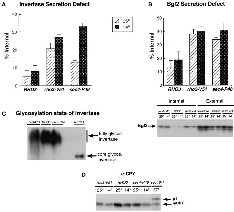
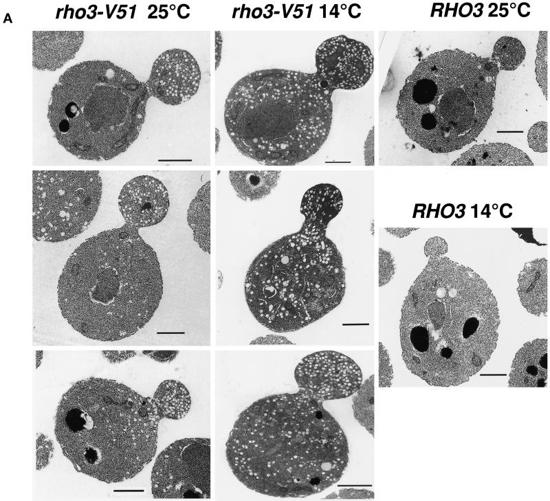
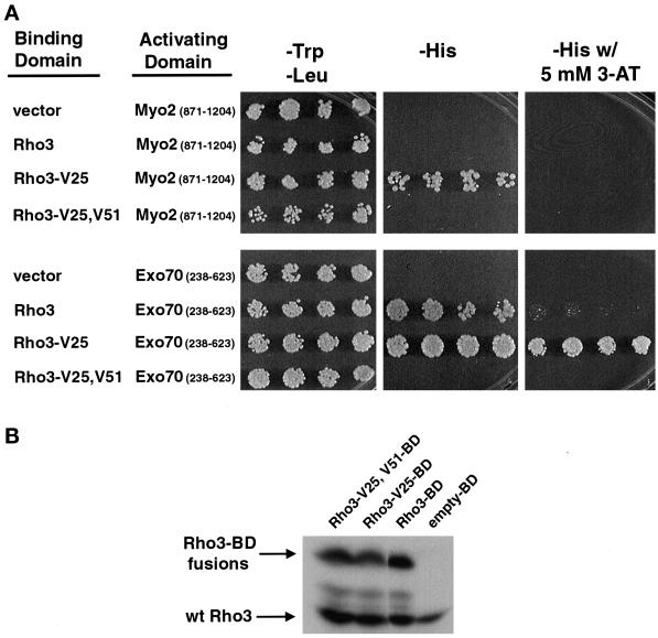
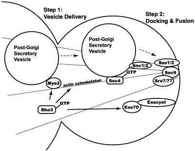
Budding yeast grow asymmetrically by the polarized delivery of proteins and lipids to specific sites on the plasma membrane. This requires the coordinated polarization of the actin cytoskeleton and the secretory apparatus. We identified Rho3 on the basis of its genetic interactions with several late-acting secretory genes. Mutational analysis of the Rho3 effector domain reveals three distinct functions in cell polarity: regulation of actin polarity, transport of exocytic vesicles from the mother cell to the bud, and docking and fusion of vesicles with the plasma membrane. We provide evidence that the vesicle delivery function of Rho3 is mediated by the unconventional myosin Myo2 and that the docking and fusion function is mediated by the exocyst component Exo70. These data suggest that Rho3 acts as a key regulator of cell polarity and exocytosis, coordinating several distinct events for delivery of proteins to specific sites on the cell surface.


Figures

Figure 1
Figure 1
HSS43 is allelic to RHO3. (A) A restriction map of the 10-kb HSS43-suppressing fragment and the subclones derived from it are shown. (a) HSS43 (10 kb). (b) PvuII fragment. (c) SalI fragment. (d) SalI-SmaI fragment. (e) SalI-XhoI 2.2-kb fragment. (B) The SalI-XhoI fragment containing the entire RHO3 open reading frame (ORF) is exclusively responsible for the suppression seen by HSS43.
Figure 2
Figure 2
RHO3 is a specific suppressor of sec4-P48, and the GTP-bound form is the active form. (A) RHO3 is the only one of the five RHO genes, RHO1, RHO2, RHO3, RHO4, or CDC42, that can suppress the cold sensitivity of sec4-P48. (B) Activated alleles of RHO3 suppress at a single copy. RHO3-V25 has amino acid 25 changed from a glycine to a valine that is analogous to the ras-V12-activating mutation. RHO3-N30 has the threonine at amino acid 30 changed to an asparagine, analogous to the ras-N17 dominant-inhibitory mutation. Each of these was introduced into sec4-P48 at a single copy, and transformants were tested for suppression of the cold sensitivity.
Figure 3
Figure 3
Mutagenesis of the Rho3 effector domain. (A) Alignment of the 14 amino acids of the effector domains of Rho3, H-ras, and Cdc42. (B) Forty-six single-point mutations generated by oligo-directed mutagenesis. Growth at each temperature was recorded as follows: (+) = wild-type growth, (+/−) = borderline growth, (−/+) = slight growth above background, and (−) = no growth. (C) Growth of the rho3 mutants at the permissive and restrictive temperature. Plates at 25°C were incubated for 4 d, and plates at 14°C plates were incubated for 7 d.
Figure 4
Figure 4
Actin localization in wild-type and rho3 mutant cells. Cells were grown at 25°C or shifted to 14°C for 5 h before fixation. Fixed cells were subsequently permeabilized and stained as described in MATERIALS AND METHODS. Bar, 5 μm.
Figure 5
Figure 5
The rho3-V51 mutant is defective in the secretion of the periplasmic enzymes invertase and Bgl2. (A) Invertase enzyme assays show a defect in the secretion for rho3-V51 at both the permissive and restrictive temperature. Secretion assays are presented as a percentage of invertase that is found internally. This is a positive measure of the secretory defect associated with a particular strain. This percentage of internal invertase is calculated by internal/(internal + external), where these values are measured as micromoles per minute per OD599. The data represent the average ± SD for five assays. (B) Immunoblot analysis of Bgl2 protein distribution shows a defect in secretion for rho3-V51 at both the permissive and restrictive temperature. The distribution of the secreted protein Bgl2 was determined by immunoblot analysis using affinity-purified antibodies raised against the C terminus of Bgl2. The band corresponding to Bgl2 is indicated. Top, quantitation of the bands was done using ImageQuant software (Molecular Dynamics, Sunnyvale, CA). The results are presented as a percentage of the total Bgl2 that is found internally. The data represent the average ± SD for three experiments. Bottom, a representative Bgl2 blot is shown. (C) Immunoblot analysis of invertase reveals the glycosylation state of invertase in these cells. (D) Transport of the vacuolar protein CPY is not affected in rho3-V51, because it is found exclusively in the mature form (mCPY, 61 kDa) at both temperatures. A sec18-1 mutant was included as a size marker and exhibits accumulation of the core glycosylated form (p1, 67 kDa). glycos., glycosylated.
Figure 6
Figure 6
Electron microscopy of rho3-V51 cells reveals accumulation of post-Golgi vesicles and a conditional defect in vesicle delivery from the mother to the bud. (A) Three panels of rho3-V51 at both 25 and 14°C as well as single panels of RHO3 at 25 and 14°C are shown. Preparation of the cells is described in MATERIALS AND METHODS. (B) Cells were scored for the location of accumulated vesicles, and bar graphs show the percentage of cells with vesicles in the bud/neck region, in the mother cell, and in the entire cell. Approximately 30 cells were counted for rho3-V51 at each temperature, and 20 cells were counted for the RHO3 cells (wt). Only small budded cells were counted. Bars, 1 μm.
Figure 6
Figure 6
Electron microscopy of rho3-V51 cells reveals accumulation of post-Golgi vesicles and a conditional defect in vesicle delivery from the mother to the bud. (A) Three panels of rho3-V51 at both 25 and 14°C as well as single panels of RHO3 at 25 and 14°C are shown. Preparation of the cells is described in MATERIALS AND METHODS. (B) Cells were scored for the location of accumulated vesicles, and bar graphs show the percentage of cells with vesicles in the bud/neck region, in the mother cell, and in the entire cell. Approximately 30 cells were counted for rho3-V51 at each temperature, and 20 cells were counted for the RHO3 cells (wt). Only small budded cells were counted. Bars, 1 μm.
Figure 7
Figure 7
Two-hybrid analysis of the effect of the V51 mutation on the interaction of Rho3 with Myo2 and Exo70. (A) Two-hybrid analysis of Rho3, Rho3-V25, and Rho3-V25,V51 mutants on the interaction with Myo2 and Exo70. Constructs containing GAL4BD-RHO3 and the GAL4AD-MYO2 (encoding residues 871-1024) or EXO70-GAL4AD (encoding residues 238–623) were transformed into PJ694α. Four independent transformants were replicated onto media selecting for both plasmids (−Trp, −Leu) or media that require activation of the HIS3 reporter (−His), and interactions were assayed by the ability of the two GAL4 fusions to activate the reporter gene HIS3. Growth on media lacking histidine or on media lacking histidine with 5 mM 3-aminotriazole (3-AT), which functions as an inhibitor of the histidine biosynthetic pathway and requires a higher level of HIS3 activation to support growth, is shown. (B) Western blot analysis of Rho3 protein levels in the two-hybrid transformants using affinity-purified α-Rho3 antibodies. Cotransformants were examined for the expression of the Gal4-binding domain (BD) fusion proteins. The endogenous Rho3 protein migrates at ∼29 kDa, and a larger fusion protein of the expected size is visible migrating at ∼48 kDa.
Figure 8
Figure 8
A model for the involvement of Rho3 in two steps of exocytosis. GTP-bound Rho3 appears to have three distinct effector pathways. Previous work (Matsui and Toh-e, 1992b; Imai et al., 1996) and results presented here show a role for Rho3 in regulating the polarized distribution of the actin cytoskeleton. Here we demonstrate a role for Rho3 in exocytosis that is independent of its effect on the actin cytoskeleton. In particular, genetic and electron microscopy data suggest that Rho3 acts both in the delivery of Golgi-derived vesicles from the mother to the bud (Step 1) and in the docking and fusion of these vesicles with the plasma membrane (Step 2). The data presented here provide strong support for the model suggested by the two-hybrid interactions identified by Robinson et al. (1999). Therefore our analysis of the rho3-V51 effector domain mutant supports the model that the vesicle delivery function of Rho3 (Step 1) is mediated by Myo2 and that the vesicle docking and fusion function of Rho3 (Step 2) is mediated by Exo70.

References

    1. Aalto MK, Ronne H, Keranen S. Yeast syntaxins Sso1p and Sso2p belong to a family of related membrane proteins that function in vesicular transport. EMBO J. 1993;12:4095–4104. - PMC - PubMed
    1. Aspenström P. Effectors for the Rho GTPases. Curr Opin Cell Biol. 1999;11:95–102. - PubMed
    1. Bourne HR, Sanders DA, McCormick F. The GTPase superfamily: conserved structure and molecular mechanism. Nature. 1991;349:117–127. - PubMed
    1. Brennwald P, Kearns B, Champion K, Keranen S, Bankaitis V, Novick P. Sec9 is a SNAP-25-like component of a yeast SNARE complex that may be the effector of Sec4 function in exocytosis. Cell. 1994;79:245–258. - PubMed
    1. Chant J, Pringle JR. Budding and cell polarity in Saccharomyces cerevisiae. Curr Opin Genet Dev. 1991;3:342–350. - PubMed

Publication types

MeSH terms