. 1999 Dec;10(12):4177-90.
doi: 10.1091/mbc.10.12.4177.

Cell-matrix adhesions differentially regulate fascin phosphorylation

Affiliations
Free PMC article

Cell-matrix adhesions differentially regulate fascin phosphorylation

J C Adams et al. Mol Biol Cell. 1999 Dec.
Free PMC article

Abstract

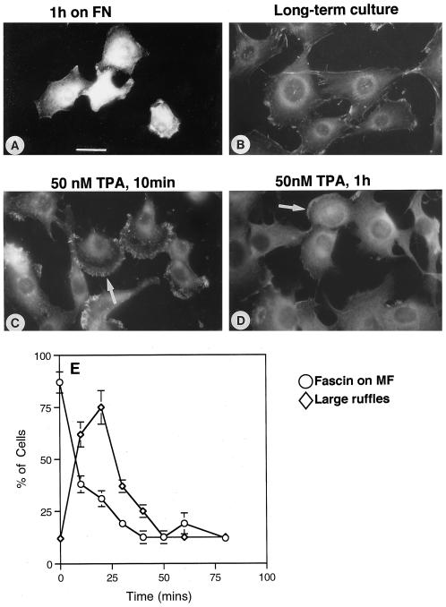
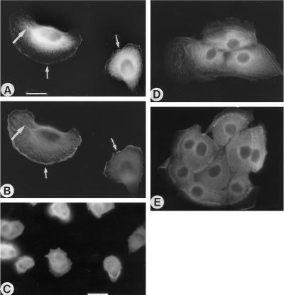
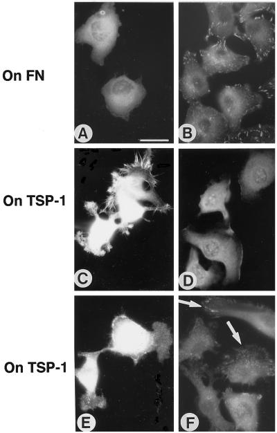
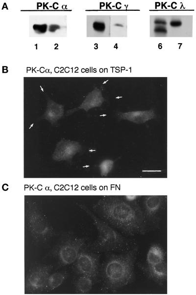
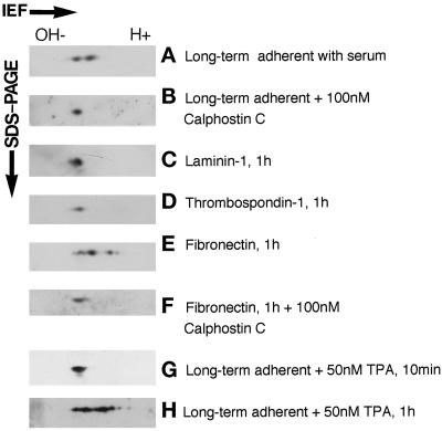
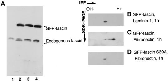
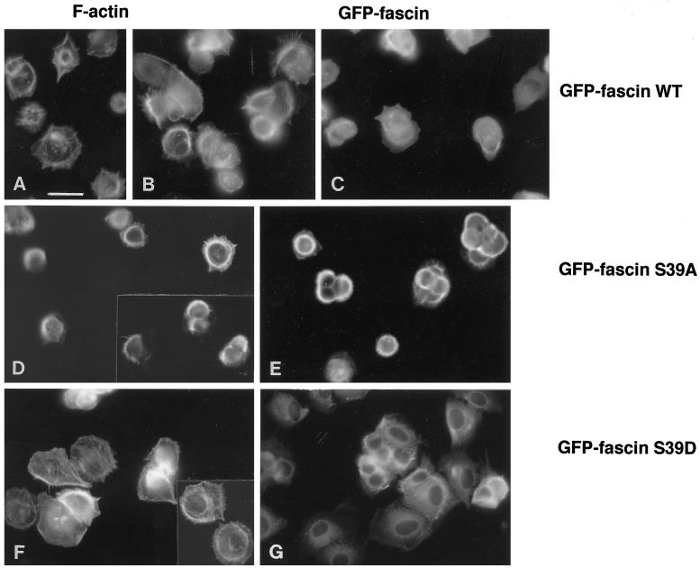
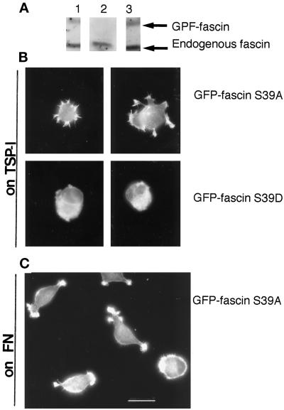
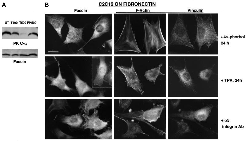
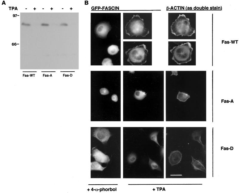
Cell adhesion to individual macromolecules of the extracellular matrix has dramatic effects on the subcellular localization of the actin-bundling protein fascin and on the ability of cells to form stable fascin microspikes. The actin-binding activity of fascin is down-regulated by phosphorylation, and we used two differentiated cell types, C2C12 skeletal myoblasts and LLC-PK1 kidney epithelial cells, to examine the hypothesis that cell adhesion to the matrix components fibronectin, laminin-1, and thrombospondin-1 differentially regulates fascin phosphorylation. In both cell types, treatment with the PKC activator 12-tetradecanoyl phorbol 13-acetate (TPA) or adhesion to fibronectin led to a diffuse distribution of fascin after 1 h. C2C12 cells contain the PKC family members alpha, gamma, and lambda, and PKCalpha localization was altered upon cell adhesion to fibronectin. Two-dimensional isoelectric focusing/SDS-polyacrylamide gels were used to determine that fascin became phosphorylated in cells adherent to fibronectin and was inhibited by the PKC inhibitors calphostin C and chelerythrine chloride. Phosphorylation of fascin was not detected in cells adherent to thrombospondin-1 or to laminin-1. LLC-PK1 cells expressing green fluorescent protein (GFP)-fascin also displayed similar regulation of fascin phosphorylation. LLC-PK1 cells expressing GFP-fascin S39A, a nonphosphorylatable mutant, did not undergo spreading and focal contact organization on fibronectin, whereas cells expressing a GFP-fascin S39D mutant with constitutive negative charge spread more extensively than wild-type cells. In contrast, C2C12 cells coexpressing S39A fascin with endogenous fascin remained competent to form microspikes on thrombospondin-1, and cells that expressed fascin S39D attached to thrombospondin-1 but did not form microspikes. Blockade of PKCalpha activity by TPA-induced down-regulation led to actin association of wild-type fascin in fibronectin-adherent C2C12 and LLC-PK1 cells but did not alter the distribution of S39A or S39D fascins. The association of fascin with actin in fibronectin-adherent cells was also evident in the presence of an inhibitory antibody to integrin alpha5 subunit. These novel results establish matrix-initiated PKC-dependent regulation of fascin phosphorylation at serine 39 as a mechanism whereby matrix adhesion is coupled to the organization of cytoskeletal structure.


Figures

Figure 1
Figure 1
Fibronectin adhesion and TPA treatment alter fascin localization in C2C12 cells. C2C12 cells were stained for fascin after 1 h of adhesion on fibronectin in serum-free medium (A), in long-term culture 1 h after addition of DMSO solvent control (B), after 50 nM TPA for 10 min (C), and after 50 nM TPA for 1 h (D). An example of a large ruffle is arrowed in C, and the residue of a ruffle is arrowed in D. Bar, 10 μm. (E) The percentage of cells with microfilament-associated fascin (○) or large ruffles (⋄) was quantitated with time of TPA treatment. Each point is the mean ± SEM of four independent experiments.
Figure 2
Figure 2
Effects of matrix adhesion or TPA treatment on GFP-fascin localization in LLC-PK1 cells. Cells adherent on laminin-1 for 1 h were double-stained with GFP-fascin (A) or antibody to β-actin (B). (C–E) GFP-fascin visualized after 1 h on fibronectin (C), in long-term adherent cells (D), or after treatment with 50 nM TPA for 1 h (E). Large arrows in A and B indicate coincidence of fascin with microfilaments, and small arrows indicate examples of coincidence in membrane ruffles. Bar, 5 μm for A and B, 10 μm for other panels.
Figure 3
Figure 3
Effect of PKC activation on microspikes and focal contacts in matrix-adherent C2C12 cells. (A and B) Cells adherent for 1 h on fibronectin in the presence of 50 nM TPA. (C and E) Control C2C12 cells adherent for 1 h on TSP-1. (D and F) Cells adherent on TSP-1 for 1 h in the presence of 50 nM TPA. (A, C, and D) Fascin stain. (B, E, and F) Vinculin stain. Arrows in F indicate localization of vinculin in focal contacts. Bar, 10 μm.
Figure 4
Figure 4
Expression of PKC family members in C2C12 cells and effect of matrix adhesion on PKCα localization. (A) Western blots of brain extract (lanes1, 3, and 6) and C2C12 whole cell extract (lanes 2, 4, and 7) were probed with antibodies to PKCα, PKCγ, and PKCλ. (B and C) C2C12 cells adherent for 1 h on TSP-1 (B) or fibronectin (FN) (C) were stained with antibody to PKCα. Arrows indicate regions of microspike formation in TSP-1–adherent cells that do not stain for PKCα. Bar, 8 μm.
Figure 5
Figure 5
Characterization of anti-fascin serum. Western blots of C2C12 whole cell extracts probed with preimmune or FAS-C serum in the presence or absence of FAS-C peptide, as indicated. PM serum, anti-fascin reagent of Pierre McCrea.
Figure 6
Figure 6
Matrix adhesion molecules differentially affect fascin phosphorylation. Whole cell extracts of C2C12 cells prepared under different adhesion conditions, as indicated, were resolved in two dimensions by IEF and SDS-PAGE, blotted, and probed with FAS-C IgG.
Figure 7
Figure 7
Requirement for serine 39 in fibronectin-dependent phosphorylation of fascin. (A) Western blot of LLC-PK1 lines probed with FAS-C serum. (Lane 1) Parental cells; (lane 2) GFP-fascin overexpressors; (lane 3) GFP-fascin S39A overexpressors; (lane 4) GFP-fascin S39D overexpressors. (B–D) Western blots of two-dimensional IEF/SDS-PAGE gels of LLC-PK1 whole cell extracts probed with FAS-C serum. (B) LLC-PK1 GFP-fascin overexpressors after 1 h on laminin-1. (C) LLC-PK1 GFP-fascin overexpressors after 1 h on fibronectin. (D) LLC-PK1 GFP-fascin S39A overexpressors after 1 h on fibronectin.
Figure 8
Figure 8
Mutant fascins affect cytoskeletal organization in fibronectin-adherent LLC-PK1 cells. Cells expressing GFP-fascin (A–C), GFP-fascin S39A (D and E), or GFP-fascin S39D (F and G) were stained with TRITC-phalloidin (A, B, D, and F) or fixed to visualize GFP-fascin (C, E, and G) after 1 h of adhesion to fibronectin. Bar, 10 μm.
Figure 9
Figure 9
Mutant fascins affect cytoskeletal organization in fibronectin-adherent C2C12 cells. (A) Western blots of whole cell extracts of C2C12 stably expressing GFP-fascin S39A (lane 1), C2C12 (lane 2), or GFP-fascin S39D (lane 3). (B) F-actin organization in C2C12 cells expressing GFP-fascin S39A or S39D after 1 h of adhesion to TSP-1. (C) F-actin organization in C2C12 cells expressing GFP-fascin S39A after 1 h of adhesion to fibronectin. Bar, 10 μm.
Figure 10
Figure 10
Requirement for PKCα activity in matrix-dependent fascin localization in C2C12 cells. (A) Western blot of C2C12 whole cell extracts probed with antiserum to PKCα or FAS-C to fascin. (UT) Untreated cells; (T100) cells treated with 100 nM TPA for 24 h; (T500) cells treated with 500 nM TPA for 24 h; (PH500) cells treated with 500 nM 4-α-phorbol for 24 h. (B) Comparison of the localizations of fascin, F-actin, and vinculin in cells treated with 500 nM 4-α-phorbol, 500 nM TPA, or 5 μg/ml 5H10 antibody to integrin α5 subunit after 1 h of adhesion to fibronectin under serum-free conditions. Bar, 5 μm.
Figure 11
Figure 11
Requirements for PKCα activity and fascin phosphorylation at serine 39 in matrix-dependent fascin localization in LLC-PK1 cells. (A) Western blot of whole cell extracts of LLC-PK1 cell lines expressing GFP-fascin (Fas-WT), GFP-S39A fascin (Fas-A), or GFP-S39D fascin (Fas-D) prepared after 24 h of treatment with 100 nM 4-α-phorbol (TPA −) or 100 nM TPA (TPA +). The blot was probed with antiserum to PKCα. (B) Comparison of GFP-fascin localization in cells treated with 100 nM 4-α-phorbol or TPA for 24 h after 1 h of adhesion to fibronectin under serum-free conditions. TPA-treated cells are shown double stained for β-actin. Bar, 5 μm.

Similar articles

Cited by

References

    1. Adams JC. Formation of stable microspikes containing actin and the 55 kDa actin-bundling protein, fascin, is a consequence of cell adhesion to thrombospondin-1: implications for the antiadhesive activities of thrombospondin-1. J Cell Sci. 1995;108:1977–1990. - PubMed
    1. Adams JC. Characterization of cell-matrix adhesion requirements for the formation of fascin microspikes. Mol Biol Cell. 1997;8:2345–2363. - PMC - PubMed
    1. Adams JC, Lawler J. Cell-type specific adhesive interactions of skeletal myoblasts with thrombospondin-1. Mol Biol Cell. 1994;5:623–637. - PMC - PubMed
    1. Adams JC, Seed B, Lawler J. Muskelin, a novel intracellular mediator of cell adhesive and cytoskeletal responses to thrombospondin-1. EMBO J. 1998;17:4964–4974. - PMC - PubMed
    1. Adams JC, Watt FM. Regulation of development and differentiation by the extracellular matrix. Development. 1993;117:1183–1198. - PubMed

Publication types

MeSH terms

LinkOut - more resources