

Initiation of assembly of the cell envelope barrier structure of stratified squamous epithelia
- PMID: 10588656
- PMCID: PMC25756
- DOI: 10.1091/mbc.10.12.4247
Initiation of assembly of the cell envelope barrier structure of stratified squamous epithelia
Abstract
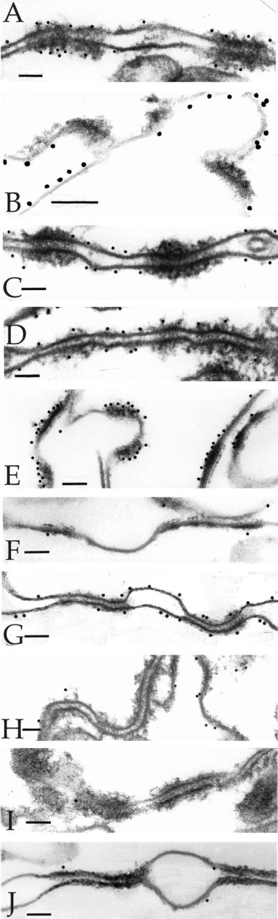
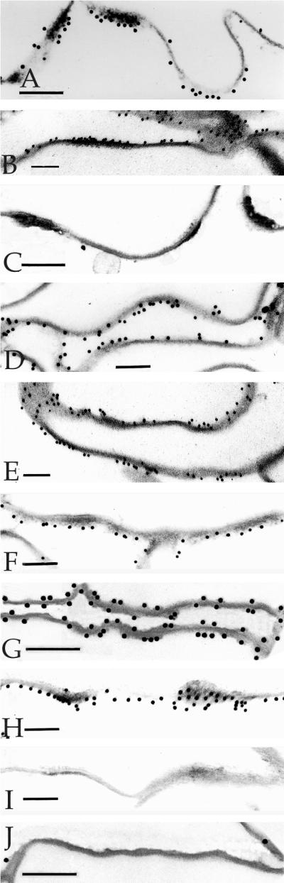
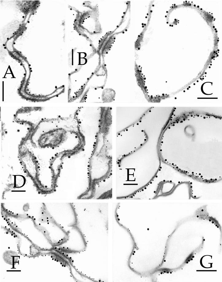
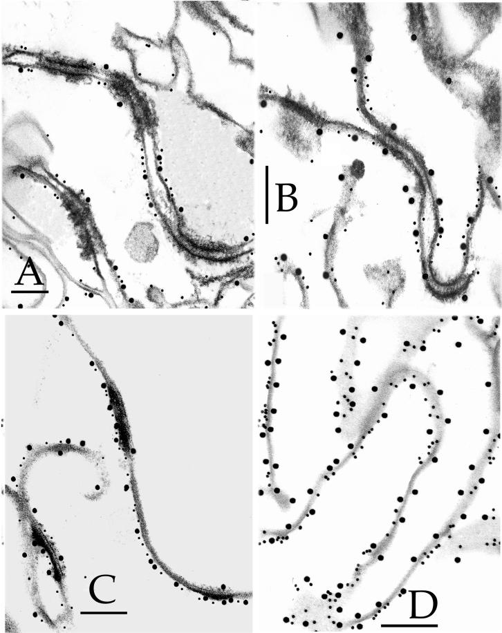
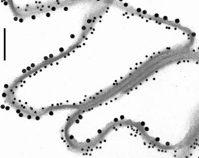
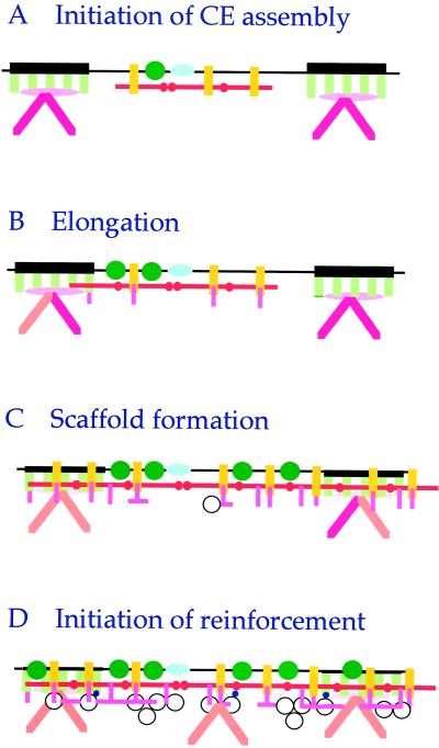
The cell envelope (CE) is a specialized structure that is important for barrier function in terminally differentiated stratified squamous epithelia. The CE is formed inside the plasma membrane and becomes insoluble as a result of cross-linking of constituent proteins by isopeptide bonds formed by transglutaminases. To investigate the earliest stages of assembly of the CE, we have studied human epidermal keratinocytes induced to terminally differentiate in submerged liquid culture as a model system for epithelia in general. CEs were harvested from 2-, 3-, 5-, or 7-d cultured cells and examined by 1) immunogold electron microscopy using antibodies to known CE or other junctional proteins and 2) amino acid sequencing of cross-linked peptides derived by proteolysis of CEs. Our data document that CE assembly is initiated along the plasma membrane between desmosomes by head-to-tail and head-to-head cross-linking of involucrin to itself and to envoplakin and perhaps periplakin. Essentially only one lysine and two glutamine residues of involucrin and two glutamines of envoplakin were used initially. In CEs of 3-d cultured cells, involucrin, envoplakin, and small proline-rich proteins were physically located at desmosomes and had become cross-linked to desmoplakin, and in 5-d CEs, these three proteins had formed a continuous layer extending uniformly along the cell periphery. By this time >15 residues of involucrin were used for cross-linking. The CEs of 7-d cells contain significant amounts of the protein loricrin, typically expressed at a later stage of CE assembly. Together, these data stress the importance of juxtaposition of membranes, transglutaminases, and involucrin and envoplakin in the initiation of CE assembly of stratified squamous epithelia.
Figures
References
-
- Akiyama M, Smith LT, Yoneda K, Holbrook KA, Hohl D, Shimizu H. Periderm cells form cornified cell envelope in their regression process during human epidermal development. J Invest Dermatol. 1999;112:903–909. - PubMed
-
- An G, Tesfaigzi J, Chuu Y-J, Wu R. Isolation and characterization of the human spr1 gene and its regulation of expression by phorbol ester and cyclic AMP. J Biol Chem. 1993;268:10977–10982. - PubMed
-
- Candi E, Lahm A, Ceci R, Rossi A, Kim I-G, Ciani B, Melino G, Steinert PM. Transglutaminase 1 mutations in lamellar ichthyosis: loss of activity due to failure of activation by proteolytic processing. J Biol Chem. 1998a;273:13693–13702. - PubMed
-
- Candi E, Melino G, Pei G, Tarcsa E, Marekov LN, Steinert PM. Bacterially-expressed human loricrin: biochemical, structural and transglutaminase substrate properties of the major epidermal cornified cell envelope structural protein. J Biol Chem. 1995;270:26382–26390. - PubMed
-
- Candi E, Tarcsa E, DiGiovanna JJ, Compton JG, Elias PM, Marekov LN, Steinert PM. A highly conserved lysine residue on the head domain of type II keratins is essential for the attachment of keratin intermediate filaments to the cornified cell envelope through isopeptide crosslinking by transglutaminases. Proc Natl Acad Sci USA. 1998b;95:2067–2072. - PMC - PubMed
MeSH terms
Substances
LinkOut - more resources
Full Text Sources
Other Literature Sources
Molecular Biology Databases
Miscellaneous
