

Correlation of the structural and functional domains in the membrane protein Vpu from HIV-1
- PMID: 10588706
- PMCID: PMC24437
- DOI: 10.1073/pnas.96.25.14336
Correlation of the structural and functional domains in the membrane protein Vpu from HIV-1
Abstract
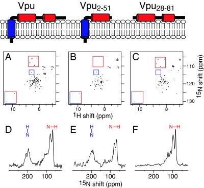
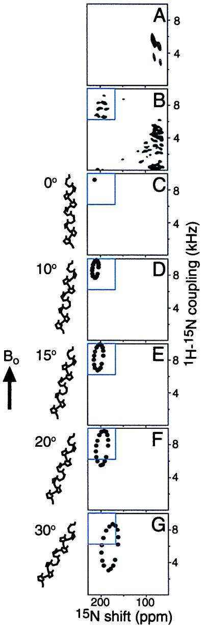
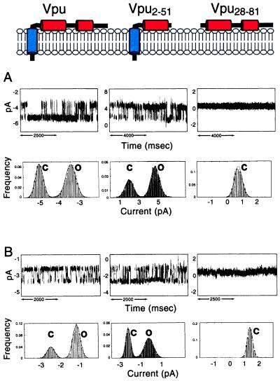
Vpu is an 81-residue membrane protein encoded by the HIV-1 genome. NMR experiments show that the protein folds into two distinct domains, a transmembrane hydrophobic helix and a cytoplasmic domain with two in-plane amphipathic alpha-helices separated by a linker region. Resonances in one-dimensional solid-state NMR spectra of uniformly (15)N labeled Vpu are clearly segregated into two bands at chemical shift frequencies associated with NH bonds in a transmembrane alpha-helix, perpendicular to the membrane surface, and with NH bonds in the cytoplasmic helices parallel to the membrane surface. Solid-state NMR spectra of truncated Vpu(2-51) (residues 2-51), which contains the transmembrane alpha-helix and the first amphipathic helix of the cytoplasmic domain, and of a construct Vpu(28-81) (residues 28-81), which contains only the cytoplasmic domain, support this structural model of Vpu in the membrane. Full-length Vpu (residues 2-81) forms discrete ion-conducting channels of heterogeneous conductance in lipid bilayers. The most frequent conductances were 22 +/- 3 pS and 12 +/- 3 pS in 0.5 M KCl and 29 +/- 3 pS and 12 +/- 3 pS in 0.5 M NaCl. In agreement with the structural model, truncated Vpu(2-51), which has the transmembrane helix, forms discrete channels in lipid bilayers, whereas the cytoplasmic domain Vpu(28-81), which lacks the transmembrane helix, does not. This finding shows that the channel activity is associated with the transmembrane helical domain. The pattern of channel activity is characteristic of the self-assembly of conductive oligomers in the membrane and is compatible with the structural and functional findings.
Figures
Similar articles
-
Expression, purification, and activities of full-length and truncated versions of the integral membrane protein Vpu from HIV-1.Protein Sci. 2002 Mar;11(3):546-57. doi: 10.1110/ps.37302. Protein Sci. 2002. PMID: 11847278 Free PMC article.
-
Three-dimensional structure of the channel-forming trans-membrane domain of virus protein "u" (Vpu) from HIV-1.J Mol Biol. 2003 Oct 17;333(2):409-24. doi: 10.1016/j.jmb.2003.08.048. J Mol Biol. 2003. PMID: 14529626
-
Three-dimensional structure of the transmembrane domain of Vpu from HIV-1 in aligned phospholipid bicelles.Biophys J. 2006 Oct 15;91(8):3032-42. doi: 10.1529/biophysj.106.087106. Epub 2006 Jul 21. Biophys J. 2006. PMID: 16861273 Free PMC article.
-
Structure-function correlates of Vpu, a membrane protein of HIV-1.FEBS Lett. 2003 Sep 18;552(1):47-53. doi: 10.1016/s0014-5793(03)00849-4. FEBS Lett. 2003. PMID: 12972151 Review.
-
Comparative NMR studies demonstrate profound differences between two viroporins: p7 of HCV and Vpu of HIV-1.Biochim Biophys Acta. 2011 Feb;1808(2):554-60. doi: 10.1016/j.bbamem.2010.08.005. Epub 2010 Aug 18. Biochim Biophys Acta. 2011. PMID: 20727848 Free PMC article. Review.
Cited by
-
Relating structure and function of viral membrane-spanning miniproteins.Curr Opin Virol. 2015 Jun;12:121-5. doi: 10.1016/j.coviro.2015.05.006. Epub 2015 Jun 6. Curr Opin Virol. 2015. PMID: 26057606 Free PMC article. Review.
-
Conformational changes induced by a single amino acid substitution in the trans-membrane domain of Vpu: implications for HIV-1 susceptibility to channel blocking drugs.Protein Sci. 2007 Oct;16(10):2205-15. doi: 10.1110/ps.073041107. Epub 2007 Aug 31. Protein Sci. 2007. PMID: 17766368 Free PMC article.
-
Modulation of beta-catenin and E-cadherin interaction by Vpu increases human immunodeficiency virus type 1 particle release.J Virol. 2008 Apr;82(8):3932-8. doi: 10.1128/JVI.00430-07. Epub 2008 Feb 6. J Virol. 2008. PMID: 18256147 Free PMC article.
-
Large multiple transmembrane domain fragments of a G protein-coupled receptor: biosynthesis, purification, and biophysical studies.Biopolymers. 2012;98(5):485-500. doi: 10.1002/bip.22122. Biopolymers. 2012. PMID: 23203693 Free PMC article.
-
Functional domains within the human immunodeficiency virus type 2 envelope protein required to enhance virus production.J Virol. 2005 Mar;79(6):3627-38. doi: 10.1128/JVI.79.6.3627-3638.2005. J Virol. 2005. PMID: 15731257 Free PMC article.
References
-
- Strebel K, Klimkait T, Martin M A. Science. 1988;241:1221–1223. - PubMed
-
- Cohen E, Terwilliger E F, Sordroski J G, Haseltine W A. Nature (London) 1988;334:532–534. - PubMed
-
- Strebel K. In: Human Retroviruses and AIDS 1996: A Compilation and Analysis of Nucleic Acid and Amino Acid Sequences. Myers G, Korber B T, Foley B T, Jeang K-T, Mellors J W, Wain-Hobson S, editors. Los Alamos, NM: Los Alamos Natl. Lab.; 1996. pp. 19–27.
Publication types
MeSH terms
Substances
Grants and funding
LinkOut - more resources
Full Text Sources
