. 1999 Dec 7;96(25):14412-7.
doi: 10.1073/pnas.96.25.14412.

The DNMT3B DNA methyltransferase gene is mutated in the ICF immunodeficiency syndrome

Affiliations

The DNMT3B DNA methyltransferase gene is mutated in the ICF immunodeficiency syndrome

R S Hansen et al. Proc Natl Acad Sci U S A. .

Abstract

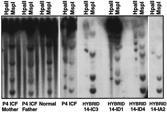
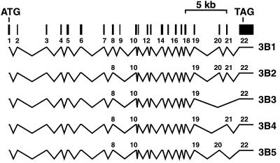
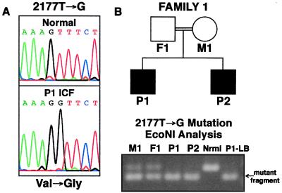
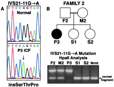
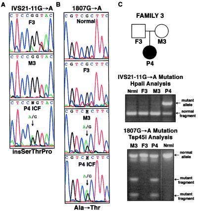
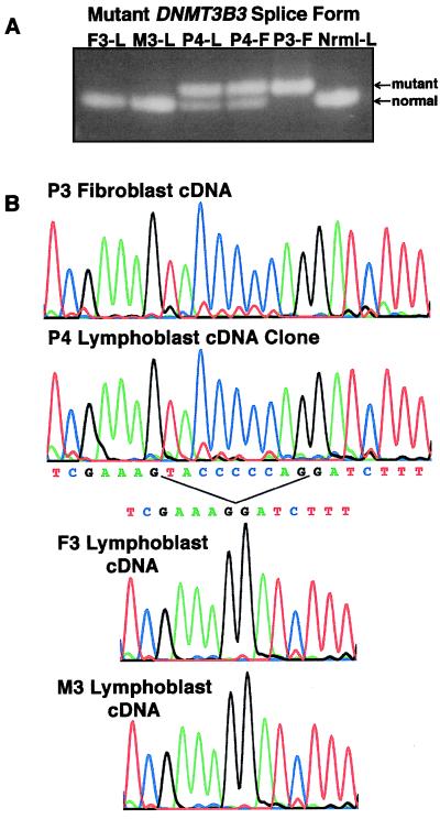
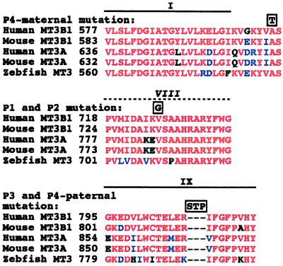
DNA methylation is an important regulator of genetic information in species ranging from bacteria to humans. DNA methylation appears to be critical for mammalian development because mice nullizygous for a targeted disruption of the DNMT1 DNA methyltransferase die at an early embryonic stage. No DNA methyltransferase mutations have been reported in humans until now. We describe here the first example of naturally occurring mutations in a mammalian DNA methyltransferase gene. These mutations occur in patients with a rare autosomal recessive disorder, which is termed the ICF syndrome, for immunodeficiency, centromeric instability, and facial anomalies. Centromeric instability of chromosomes 1, 9, and 16 is associated with abnormal hypomethylation of CpG sites in their pericentromeric satellite regions. We are able to complement this hypomethylation defect by somatic cell fusion to Chinese hamster ovary cells, suggesting that the ICF gene is conserved in the hamster and promotes de novo methylation. ICF has been localized to a 9-centimorgan region of chromosome 20 by homozygosity mapping. By searching for homologies to known DNA methyltransferases, we identified a genomic sequence in the ICF region that contains the homologue of the mouse Dnmt3b methyltransferase gene. Using the human sequence to screen ICF kindreds, we discovered mutations in four patients from three families. Mutations include two missense substitutions and a 3-aa insertion resulting from the creation of a novel 3' splice acceptor. None of the mutations were found in over 200 normal chromosomes. We conclude that mutations in the DNMT3B are responsible for the ICF syndrome.


Figures

Figure 1
Figure 1
Complementation of the ICF satellite hypomethylation defect after somatic cell hybridization. Satellite 2 sequences were examined for methylation status in the P4 ICF family and in somatic cell hybrid clones derived from fusion of P4 lymphoblasts to CHO cells. DNAs were digested with either the methylation-sensitive enzyme HpaII or its methylation-insensitive isoschizimer, MspI. In normal lymphoblasts, the satellite sequences are resistant to HpaII digestion whereas MspI digestion results in a ladder of fragments. This pattern indicates that these satellites are normally hypermethylated at HpaII sites. The ICF HpaII digestion patterns, in contrast, are very similar to those of MspI, indicating that the satellites are hypomethylated. All seven somatic cell hybrid clones derived from P4 lymphoblasts that contained human satellite 2 sequences were more resistant to HpaII digestion than in the ICF patient (14-IC3, 14-ID1, 14-ID4, and 14-IA2 are shown), indicating that they had become de novo methylated in the CHO cell background.
Figure 2
Figure 2
Structure of the human DNMT3B gene. Genomic sequence, protein homology to the murine Dnmt3b gene, and cDNA sequence from RT-PCR analysis were used to determine the genomic structure of the human DNMT3B gene. Alternative splice forms were detected by RT-PCR and are depicted according to data obtained from mouse (16) and human (23, 25) cDNA clones. The exon–intron boundaries that we determined agree with those recently described by Xie et al. (23). Splice forms were named according to Robertson et al. (25).
Figure 3
Figure 3
Homozygous DNMT3B mutation in ICF Family 1. Family 1 is a consanguineous Dutch kindred with two affected brothers (P1 and P2) that was described previously (8, 40). (A) A homozygous T to G transversion in codon 726 (2177T → G) was detected in the cDNA of P1 (mt3b-14f:15r product) that changes valine to glycine. (B) An assay was developed to rapidly screen human populations for this mutation. Digestion of the 3bx19 gt-f:mt3b-20r PCR product with EcoNI only occurs with the mutant allele, as seen for Family 1 members in which the father (F1) and mother (M1) are heterozygous and the two affected children (P1 and P2) are homozygous for the mutation (DNAs were derived from blood cells, except P1-LB, which is from a lymphoblastoid cell line used in screening cDNA for mutations; nrml is a normal control).
Figure 4
Figure 4
Homozygous DNMT3B mutation in ICF Family 2. Family 2 is a consanguineous Turkish kindred with an affected female proband (P3) described by Wijmenga et al. (8). In P3 of Family 2, a homozygous CpG to CpA transition (IVS21–11G → A) was detected 11 nucleotides upstream from the normal 5′ splice acceptor site for exon 22 in the mt3b-23f:23r PCR product. The mutation can be detected by HpaII analysis of the product; normal alleles are digested by HpaII, but the mutant one is not. The father (F2) and mother (M2) of P3 are both heterozygous for the mutation and the two unaffected daughters homozygous for normal alleles. The mutation creates an AG dinucleotide that is predicted to create a new splice acceptor site that retains nine intronic nucleotides at the 5′ end of exon 22.
Figure 5
Figure 5
Heterozygous DNMT3B mutations in ICF Family 3. Family 3 is a North American kindred with a female proband (P4) and no evidence of consanguinity. (A) The IVS21–11G → A mutation described in Fig. 4 for Family 2 was also found on one P4 allele in the mt3b-23f:23r PCR product. (B) A CpG to CpA transition in codon 603 (1807G → A) was detected on one P4 allele (mt3b-11f:11r RT-PCR product) that changes alanine to threonine. This mutation is present in the mother (M3) but not the father (F3). (C) The IVS21–11G → A mutation was analyzed by HpaII digestion as described in Fig. 4. The 1807G → A mutation can be detected by the ability of Tsp45I to digest the mt3b-19f:19r PCR product; normal products are not digested.
Figure 6
Figure 6
DNMT3B alternative splice variants and sequence analysis of the exon 22 splice mutation. Amplification of cDNA with several different primer sets (mt3b-8f:8r, m3b-13f:13r, mt3b-14f:14r, mt3b-15f:15r, mt3b-16f:16r) suggested that several alternative splice forms of DNMT3B are present in normal and ICF cells. The predominant splice variant detected in both lymphoblasts (L) and fibroblasts (F) lacks exons 9, 20, and 21 (the 3B3 isoform). (A) The IVS21–11G→A mutation causes an exon 22 splice alteration. The mt3b-14f:14r RT-PCR products in P3 and P4 cDNA contain a larger 3B3 product than normal, correlating with the presence of the intron 21 mutation. (B) A 9-nt addition to exon 22 in P3 and P4 cDNA. Sequence analyses of RT-PCR products corresponding to 3B3 (mt3b-14f:14r) and 3B4 (mt3b-13f:13r; not shown) indicate that the intron 21 mutation creates a new splice acceptor site that adds nine nucleotides to the 3′ end of exon 22, effectively creating an insertion of SerThrPro just before Arg 807.
Figure 7
Figure 7
DNMT3B mutations occur at positions that are invariant in DNMT3-like methyltransferases and are within or near motifs that are conserved in most cytosine methyltransferases. DNMT3-like methyltransferases identified in zebrafish (GenBank accession no. AF135438), mice (GenBank accession nos. AF068625 for Dnmt3a and AF068626 for Dnmt3b), and humans (GenBank accession nos. AF067972 for DNMT3A and AF156488 for DNMT3B) are compared in the regions in which ICF mutations were found. Sequences were aligned with clustalw 1.7 (http://dot.imgen.bcm.tmc.edu:9331/multi-align/multi-align.html) and were analyzed by boxshade 3.21 (http://www.isrec.isb-sib.ch:8080/software/BOX_form.html). The mutations detected in the 3 ICF families are depicted above the aligned sequences in boxes. Regions that are similar to conserved DNA methyltransferase motifs are also depicted above the alignment (dotted line indicates that motif VIII similarity is weak). Key for amino acid comparisons: red, identical; blue, conservative differences; black, nonconservative differences.

References

    1. Tiepolo L, Maraschio P, Gimelli G, Cuoco C, Gargani G F, Romano C. Hum Genet. 1979;51:127–137. - PubMed
    1. Hulten M. Clin Genet. 1978;14:294.
    1. Jeanpierre M, Turleau C, Aurias A, Prieur M, Ledeist F, Fischer A, Viegas-Pequignot E. Hum Mol Genet. 1993;2:731–735. - PubMed
    1. Ji W, Hernandez R, Zhang X Y, Qu G Z, Frady A, Varela M, Ehrlich M. Mutat Res. 1997;379:33–41. - PubMed
    1. Miniou P, Jeanpierre M, Bourc’his D, Coutinho Barbosa A C, Blanquet V, Viegas-Pequignot E. Hum Genet. 1997;99:738–745. - PubMed

Publication types

Substances