. 1999 Dec 7;96(25):14505-10.
doi: 10.1073/pnas.96.25.14505.

Mouse model of Sanfilippo syndrome type B produced by targeted disruption of the gene encoding alpha-N-acetylglucosaminidase

Affiliations

Mouse model of Sanfilippo syndrome type B produced by targeted disruption of the gene encoding alpha-N-acetylglucosaminidase

H H Li et al. Proc Natl Acad Sci U S A. .

Abstract

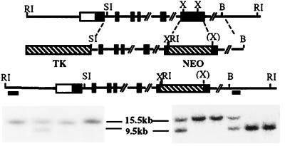
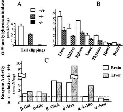
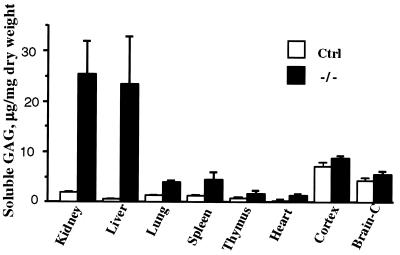
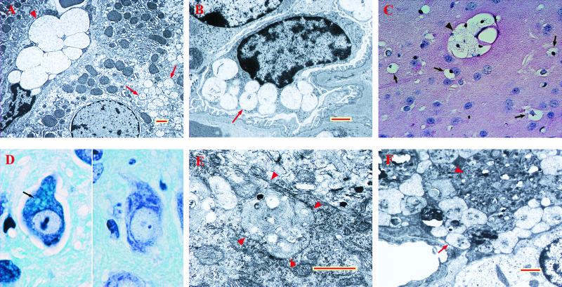
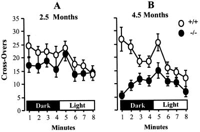
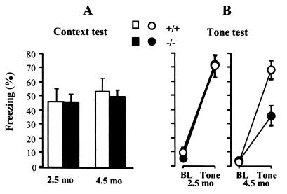
The Sanfilippo syndrome type B is an autosomal recessive disorder caused by mutation in the gene (NAGLU) encoding alpha-N-acetylglucosaminidase, a lysosomal enzyme required for the stepwise degradation of heparan sulfate. The most serious manifestations are profound mental retardation, intractable behavior problems, and death in the second decade. To generate a model for studies of pathophysiology and of potential therapy, we disrupted exon 6 of Naglu, the homologous mouse gene. Naglu-/- mice were healthy and fertile while young and could survive for 8-12 mo. They were totally deficient in alpha-N-acetylglucosaminidase and had massive accumulation of heparan sulfate in liver and kidney as well as secondary changes in activity of several other lysosomal enzymes in liver and brain and elevation of gangliosides G(M2) and G(M3) in brain. Vacuolation was seen in many cells, including macrophages, epithelial cells, and neurons, and became more prominent with age. Although most vacuoles contained finely granular material characteristic of glycosaminoglycan accumulation, large pleiomorphic inclusions were seen in some neurons and pericytes in the brain. Abnormal hypoactive behavior was manifested by 4.5-mo-old Naglu-/- mice in an open field test; the hyperactivity that is characteristic of affected children was not observed even in younger mice. In a Pavlovian fear conditioning test, the 4.5-mo-old mutant mice showed normal response to context, indicating intact hippocampal-dependent learning, but reduced response to a conditioning tone, perhaps attributable to hearing impairment. The phenotype of the alpha-N-acetylglucosaminidase-deficient mice is sufficiently similar to that of patients with the Sanfilippo syndrome type B to make these mice a good model for study of pathophysiology and for development of therapy.


Figures

Figure 1
Figure 1
Targeted disruption of the Naglu gene. The structure of the endogenous gene, the targeting construct, and the disrupted allele are presented schematically on successive lines. Southern blot identification of the disrupted gene is shown (Bottom): embryonic stem cell colonies (Left) and F2 mice (Right). These were identified by hybridization of the 5′ probe to EcoRI restriction fragments. The fragment is smaller in the disrupted allele than in the normal one (9.5 kb vs. 15.5 kb) because of the presence of an EcoRI site in the neor gene. Hybridization with the 3′ probe gave a 6-kb fragment for the disrupted allele (not shown). Filled rectangles represent exons, whereas the open rectangle at the 5′ end indicates that the transcription start site has not been defined. The two bars under the disrupted allele represent the 5′ and 3′ probes used for Southern blots. Abbreviations for restriction enzymes are: RI, EcoRI; SI, SacI; X, XhoI; B, BamHI; (X) indicates that the XhoI site was lost during the cloning process.
Figure 2
Figure 2
Enzyme activities in mutant and control mice. (A) Activity, mean ± SD, of α-N-acetylglucosaminidase in tail clippings of 10-d-old mice. The mice of the second backcross generation numbered 65 for +/+, 102 for +/−, and 65 for −/−. There was no change in enzyme level in subsequent backcross generations. (B) α-N-acetylglucosaminidase activity in various tissues of 1-mo-old +/+ and +/− mice (two mice each for brain and three for other tissues); the activity in −/− mice is not shown, because it was zero in every case. (C) Ratio of activity of other enzymes in liver and brain of +/+ and −/− mice; one 5-mo-old mutant mouse was used for liver and two for brain, with one control mouse of the same age. Abbreviations for enzymes: β-Gal, β-galactosidase; α-Glu, α-glucosidase; β-GlcA, β-glucuronidase; β-Hex, β-hexosaminidase; α-l-Idu, α-l-iduronidase; α-Neu, α-N-acetylneuraminidase.
Figure 3
Figure 3
Accumulation of soluble GAG in tissues of mutant and control mice. Soluble Alcian-blue precipitable GAG (mean ±SD) was from tissues of six −/− and eight control mice (6 +/− and 2 +/+). The brain was separated into cortex and remainder (Brain-C).
Figure 4
Figure 4
Abnormal inclusions in tissues of mutant mice. (A) Liver of −/− mouse, 3 mo old. Note the very large vacuoles in the Kupffer cell (arrowhead) and much smaller vacuoles in the hepatocyte (arrows), both containing fine granular material. (B) Podocyte of −/− mouse, 6 mo old, showing vacuoles with fine granular material (arrow). (C) Macrophage-like cells with vacuolated cytoplasm in caudatoputamen of −/− mouse, 6 mo old (hematoxylin/eosin stain). Arrows point to individual cells, whereas arrowhead points to a cluster. (D) Neuron of dentate nucleus of cerebellum with large inclusions stained light blue (arrow) in −/− mouse (Left) and absence of such inclusions in cell from same area of a control mouse, both 6 mo old (Kluver–Barrera luxol fast blue stain). (E) Purkinje cell from 6-mo-old −/− mouse. Note the large inclusion containing lamellar as well as coarse granular material; arrowheads point to edge of the inclusion. (F) Pericyte from 6-mo-old −/− mouse. A very large pleiomorphic inclusion (arrowhead) is present in addition to inclusions containing granular material (arrow). Red and yellow bars = 1 μm.
Figure 5
Figure 5
Open field behavior of mutant and control mice. Mutant (filled circles) and control +/+ mice (open circles) were tested for open field activity in darkness and in bright light. Mean ± SEM is given for crossovers scored during each minute. (A) 2.5-mo-old mice, 13 −/− and 10 +/+. (B) 4.5-mo-old mice, 9 −/− and 8 +/+.
Figure 6
Figure 6
Pavlovian fear conditioning. Mutant −/− (filled circles) and control +/+ mice (open circles), 2.5 or 4.5 mo old, were conditioned with a tone-shock pairing. Freezing is reported as % time, mean ± SEM. The mice are the same as those used in the open field test of Fig. 5. (A) Context-elicited freezing assessed for 4 min. (B) After determining baseline (BL) freezing for 2 min in a novel context, the tone was played for 4 min to assess tone-elicited freezing.

Similar articles

Cited by

References

    1. Neufeld E F, Muenzer J. In: The Metabolic and Molecular Bases of Inherited Disease. Scriver C R, Beaudet A L, Sly W S, Valle D, editors. New York: McGraw–Hill; 1995. pp. 2465–2494.
    1. Zhao H G, Li H H, Bach G, Schmidtchen A, Neufeld E F. Proc Natl Acad Sci USA. 1996;93:6101–6105. - PMC - PubMed
    1. Weber B, Blanch L, Clements P R, Scott H S, Hopwood J J. Hum Mol Genet. 1996;5:771–777. - PubMed
    1. Zhao Z, Yazdani A, Shen Y, Sun Z, Bailey J, Caskey C T, Lee C C. Mamm Genome. 1996;7:686–690. - PubMed
    1. Schmidtchen A, Greenberg D, Zhao H G, Li H H, Huang Y, Tieu P, Zhao H Z, Cheng S, Zhao Z, Whitley C B, et al. Am J Hum Genet. 1998;62:64–69. - PMC - PubMed

Publication types

Associated data