. 1999 Dec 7;96(25):14541-6.
doi: 10.1073/pnas.96.25.14541.

Prevention of lymphocyte cell death in sepsis improves survival in mice

Affiliations

Prevention of lymphocyte cell death in sepsis improves survival in mice

R S Hotchkiss et al. Proc Natl Acad Sci U S A. .

Abstract

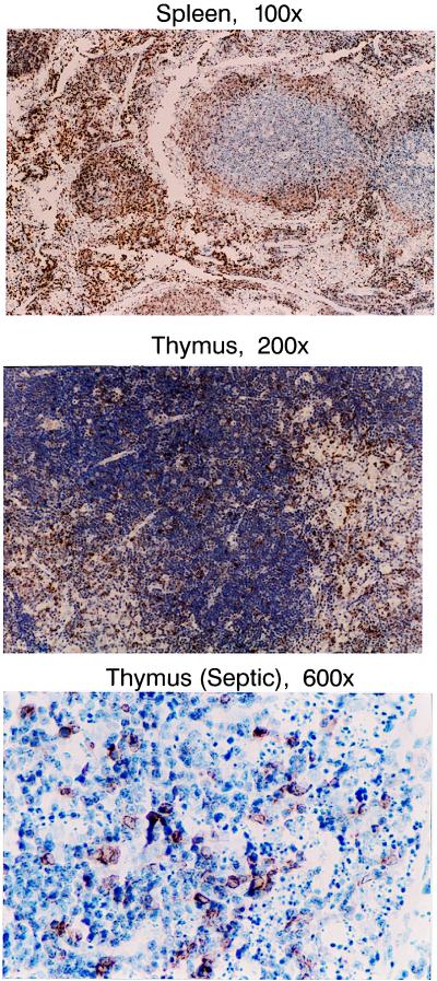
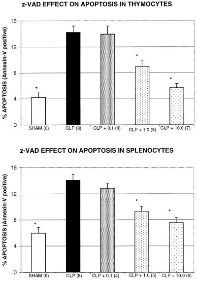
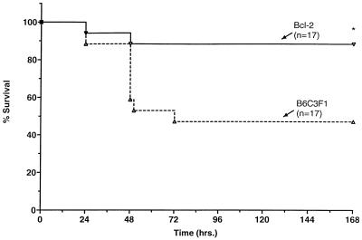
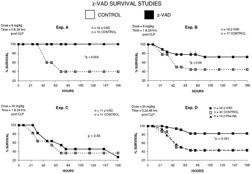
Sepsis induces extensive lymphocyte apoptosis, a process which may be beneficial to host survival by down-regulating the inflammatory response or, alternatively, harmful by impairing host defenses. To determine the beneficial vs. adverse effects of lymphocyte apoptosis in sepsis, we blocked lymphocyte apoptosis either by N-benzyloxycarbonyl-Val-Ala-Asp(O-methyl) fluoromethyl ketone (z-VAD), a broad-spectrum caspase inhibitor, or by use of Bcl-2 Ig transgenic mice that selectively overexpress the antiapoptotic protein Bcl-2 in a lymphoid pattern. Both z-VAD and Bcl-2 prevented lymphocyte apoptosis and resulted in a marked improvement in survival. z-VAD did not decrease lymphocyte tumor necrosis factor-alpha production. Considered together, these two studies employing different methods of blocking lymphocyte apoptosis provide compelling evidence that immunodepression resulting from the loss of lymphocytes is a central pathogenic event in sepsis, and they challenge the current paradigm that regards sepsis as a disorder resulting from an uncontrolled inflammatory response. Caspase inhibitors may represent a treatment strategy in this highly lethal disorder.


Figures

Figure 1
Figure 1
Bcl-2 prevents apoptosis in sepsis. Thymocytes or splenocytes from septic or sham-operated Bcl-2 transgenic or B6C3F1 mice were examined for apoptosis by flow cytometry and the apoptosis indicator annexin V. Different classes of T or B cells were determined by labeling cell markers, i.e., CD4, CD8, CD3, or CD19 with fluorophore-conjugated Abs from PharMingen. Sepsis increased apoptosis in T and B cells from thymi and spleens of B6C3F1 mice; *, P < 0.05 septic vs. sham. Although there was no protection from apoptosis in thymocytes from septic Bcl-2 transgenic mice, both T and B cells in spleens from septic Bcl-2 mice were protected against sepsis-induced apoptosis; *, P < 0.05 septic vs. sham, n = 5 sham and n = 7–8 septic for both Bcl-2 and B6C3F1 mice. The protection by Bcl-2 was consistent with the greater expression of Bcl-2 in spleen vs. thymus, as seen in immunohistochemical stains (see Fig. 2).
Figure 2
Figure 2
Immunohistochemical staining for human Bcl-2 in thymi and spleens from Bcl-2 transgenic mice. Splenocytes that have a brown color (resulting from diaminobenzidine staining) are positive for human Bcl-2. Note the much greater staining in spleen vs. thymus in the Bcl-2 transgenic mice (Top vs. Middle). Although there was much less staining in thymi vs. spleens of Bcl-2 transgenics, the cells that were positive for Bcl-2 in thymi were resistant to sepsis-induced apoptosis. As is apparent in Bottom, none of the Bcl-2-positive cells in the septic thymus were apoptotic, i.e., none of the Bcl-2-positive cells had features of pyknosis or karyorrhexis (n = 10 thymi from septic Bcl-2-Ig transgenic mice)
Figure 3
Figure 3
z-VAD prevents apoptosis in thymocytes and splenocytes during sepsis. Female ND4 mice (Harlan) underwent CLP or sham surgery, as described in Methods. One hour after surgery, mice received z-VAD at 0.1, 1.0, or 10.0 mg/kg body weight. Apoptosis was evaluated 18–22 hr after CLP or sham surgery by annexin V labeling and flow cytometry of isolated thymocytes (Upper) or splenocytes (Lower), as described in Methods. Sham-operated mice and mice treated with 1.0 or 10.0 mg/kg body weight z-VAD were significantly different from untreated CLP mice; *, P < 0.05. The number of mice per group is indicated in parentheses.
Figure 4
Figure 4
Overexpression of Bcl-2 improves survival in sepsis. Generation of the Bcl-2-Ig transgenic mice has been described previously (17) (see Methods). Bcl-2 overexpressor mice and age- and sex-matched B6C3F1 mice underwent CLP. One hour after CLP, mice received metromidazole and ceftriazone as antibiotic coverage (see Methods for details). Antibiotics were repeated every 12 hr for 48 hr, and survival was improved in Bcl-2 overexpressors vs. B6C3F1 mice; *, P < 0.02. n = number of mice in each group.
Figure 5
Figure 5
The caspase inhibitor z-VAD improves survival in sepsis. A series of survival studies were performed in which ND4 mice underwent CLP and received antibiotics, as described in Methods. Next, mice received z-VAD or the diluent in which z-VAD was dissolved. The dose (mg/kg body weight) and time of administration of z-VAD are indicated in the upper left corner of each experiment. n = number of mice per group. (Exp. A) In this study, z-VAD was administered at the same time as CLP. Mice in the z-VAD group had improved survival; *, P < 0.003. (Exp. B) Mice received z-VAD (6 mg/kg) 1 hr after CLP. Mice treated with z-VAD had improved survival at 82–84 hr after CLP, compared with control mice; *, P < 0.04. (Exp. C) Mice received high-dose z-VAD (30 mg/kg) 1 hr after CLP. There was no difference in survival in the treated vs. untreated group. (Exp. D) Three separate studies were performed, and the results were combined. Mice received 20 mg/kg z-VAD or z-FA-FMK (z-Phe-Ala; a control noncaspase inhibitor that does have limited activity to block cathepsins) 1, 24, and 48 hr after CLP. A third group of mice (CONTROL) received the diluent for the z-VAD, i.e., DMSO in PBS (see Methods). Mice in the z-VAD-treated group had improved survival compared with both z-FA-FMK (P < 0.02) and control (P < 0.001).

Similar articles

Cited by

References

    1. Stone R. Science. 1994;264:365–367. - PubMed
    1. Wang S D, Huang K J, Lin Y S, Lei H Y. J Immunol. 1994;152:5014–5021. - PubMed
    1. Ayala A, Herndon C, Lehman D, DeMaso C, Ayala C, Chaudry I. Shock. 1995;3:259–268. - PubMed
    1. Hotchkiss R S, Swanson P E, Cobb J P, Jacobson A, Buchman T G, Karl I. Crit Care Med. 1997;25:1298–1307. - PubMed
    1. Bone R C. Crit Care Med. 1996;24:1125–1129. - PubMed

Publication types

Substances