. 1999 Dec 7;96(25):14559-64.
doi: 10.1073/pnas.96.25.14559.

Altered states: involvement of phosphorylated CagA in the induction of host cellular growth changes by Helicobacter pylori

Affiliations

Altered states: involvement of phosphorylated CagA in the induction of host cellular growth changes by Helicobacter pylori

E D Segal et al. Proc Natl Acad Sci U S A. .

Abstract

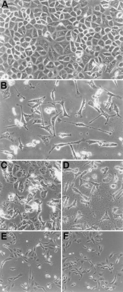
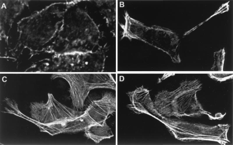
Helicobacter pylori, present in half of the world's population, is a very successful pathogen. It can survive for decades in the human stomach with few obvious consequences to the host. However, it is also the cause of gastric diseases ranging from gastritis to ulcers to gastric cancer and has been classified a type 1 carcinogen by the World Health Organization. We have previously shown that phosphorylation of a 145-kDa protein and activation of signal transduction pathways are associated with the attachment of H. pylori to gastric cells. Here we identify the 145-kDa protein as the H. pylori CagA protein. We also show that CagA is necessary to induce a growth-factor-like phenotype (hummingbird) in host gastric cells similar to that induced by hepatocyte growth factor (HGF). Additionally, we identify a second cellular phenotype induced after attachment by H. pylori, which we call SFA (stress fiber associated). SFA is CagA independent and is produced by type I and type II H. pylori.


Figures

Figure 1
Figure 1
Amino acid sequence of the CagA protein from H. pylori strain 26695 (the Institute for Genomic Research). Regions that were identified from matrix-assisted laser desorption ionization time-of-flight mass spectrometry analysis of the tyrosine-phosphorylated protein are highlighted in red.
Figure 2
Figure 2
Deconvolution immunofluorescence and 3D modeling of H. pylori attached to AGS cells. (A) H. pylori strain G27 attached to AGS cells was stained with DAPI [which stains the host-cell nucleus (large pink arrow) and bacteria (small pink arrow) nuclear DNA blue], anti-CagA (red), and antiphosphotyrosine (green). Colocalization of green and red appears yellow. The image is a composite of 0.2-μm Z-axis sections. The region indicated by the white arrow in A was used to generate the 3D wire models shown in B and C. C is a longitudinal view of B. (B and C) H. pylori is blue, CagA is red, and phosphotyrosine is green. Imaged and modeled on an Applied Precision DeltaVision Deconvolution System.
Figure 3
Figure 3
Deconvolution immunofluorescence and 3D modeling of H. pylori attached to AGS cells. H. pylori strain G27 (A–F), strain 87A300 (G–I). (A) H. pylori attached to AGS cells and stained for H. pylori (red) and CagA (green). Colocalization is yellow. The field is a 0.2-μm image. (B) H. pylori attached to AGS cells and stained for H. pylori (red) and CagA (green). Colocalization is yellow. The field is a volume composite of 0.2-μm images. (C) The region indicated by the arrow in B is magnified in C. (D) H. pylori attached to AGS cells and stained for H. pylori (blue), CagA (green), and actin (red). Colocalization of green and red appears yellow, of blue and green, cyan, and of red and blue, magenta. The field is a volume composite of 0.2-μm images. The region indicated by the arrow in D was used to generate the 3D solid models shown in E and F. F is a side view of E. (E and F) H. pylori is blue, CagA is green, and actin is red. (G) H. pylori (small pink arrow) attached to AGS cells and stained for H. pylori (blue), CagA (green), and actin (red). Colocalization of green and red appears yellow, of blue and green, cyan, and of red and blue, magenta. The field is a 0.2-μm image. The field in G was used to generate the 3D wire model shown in H. (H) H. pylori is blue, CagA is green, and actin is red. (I) H. pylori (small pink arrow) attached to AGS cells were stained with DAPI [which stains the host-cell nucleus (large pink arrow) and bacterial nuclear DNA blue], anti-CagA (green), and actin (red). Colocalization of green and red appears yellow, of blue and green, cyan, and of red and blue, magenta. The field is a volume composite of 0.2-μm images. Imaged and modeled on an Applied Precision DeltaVision Deconvolution System.
Figure 4
Figure 4
Phase contrast microscopy of AGS and MDCK cells after H. pylori attachment. (A) AGS control, 30 hr. (B) AGS plus H. pylori G27, 30 hr. (C) AGS plus H. pylori G27/cagA, 30 hr. (D) AGS plus H. pylori G27/vacA, 30 hr. (E) MDCK plus H. pylori 87A300, 24 hr. (I) MDCK plus HGF, 24 hr.
Figure 5
Figure 5
Deconvolution immunofluorescence of H. pylori attached to AGS cells for 5 hr (0.2-μm slice). All fields are stained for actin. (A) AGS control, (B) plus H. pylori G27, (C) plus H. pylori G27/cagA, (D) plus H. pylori G27/cagA/vacA. Imaged on an Applied Precision DeltaVision Deconvolution System.

Similar articles

Cited by

References

    1. Logan R P. Lancet. 1994;344:1078–1079. - PubMed
    1. Censini S, Lange C, Xiang Z, Crabtree J E, Ghiara P, Borodovsky M, Rappuoli R, Covacci A. Proc Natl Acad Sci USA. 1996;93:14648–14653. - PMC - PubMed
    1. Segal E D, Falkow S, Tompkins L S. Proc Natl Acad Sci USA. 1996;93:1259–1264. - PMC - PubMed
    1. Segal E D, Lange C, Covacci A, Tompkins L S, Falkow S. Proc Natl Acad Sci USA. 1997;94:7595–7599. - PMC - PubMed
    1. Fernandez J, Gharahdaghi F, Mische S M. Electrophoresis. 1998;19:1036–1045. - PubMed

Publication types

Substances

LinkOut - more resources