

Neuropeptide release by efficient recruitment of diffusing cytoplasmic secretory vesicles
- PMID: 10588747
- PMCID: PMC24478
- DOI: 10.1073/pnas.96.25.14577
Neuropeptide release by efficient recruitment of diffusing cytoplasmic secretory vesicles
Abstract
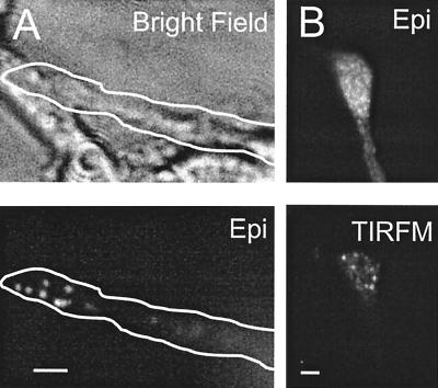
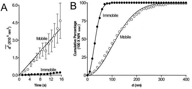
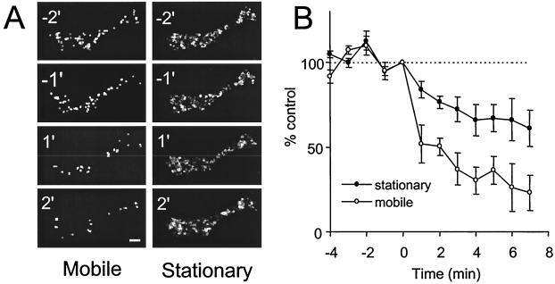
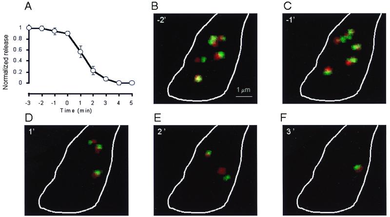
Neuropeptides are slowly released from a limited pool of secretory vesicles. Despite decades of research, the composition of this pool has remained unknown. Endocrine cell studies support the hypothesis that a population of docked vesicles supports the first minutes of hormone release. However, it has been proposed that mobile cytoplasmic vesicles dominate the releasable neuropeptide pool. Here, to determine the cellular basis of the releasable pool, single green fluorescent protein-labeled secretory vesicles were visualized in neuronal growth cones with the use of an inducible construct or total internal reflection fluorescence microscopy. We report that vesicle movement follows the diffusion equation. Furthermore, rapidly moving secretory vesicles are used more efficiently than stationary vesicles near the plasma membrane to support stimulated release. Thus, randomly moving cytoplasmic vesicles participate in the first minutes of neuropeptide release. Importantly, the preferential recruitment of diffusing cytoplasmic secretory vesicles contributes to the characteristic slow kinetics and limited extent of sustained neuropeptide release.
Figures

Similar articles
-
Neuronal peptide release is limited by secretory granule mobility.Neuron. 1997 Nov;19(5):1095-102. doi: 10.1016/s0896-6273(00)80400-6. Neuron. 1997. PMID: 9390522
-
Transport, docking and exocytosis of single secretory granules in live chromaffin cells.Nature. 1997 Jul 31;388(6641):474-8. doi: 10.1038/41329. Nature. 1997. PMID: 9242406
-
Unexpected mobility variation among individual secretory vesicles produces an apparent refractory neuropeptide pool.Biophys J. 2003 Jun;84(6):4127-34. doi: 10.1016/S0006-3495(03)75137-6. Biophys J. 2003. PMID: 12770915 Free PMC article.
-
Protein trafficking to chromaffin granules and proteolytic processing within regulated secretory vesicles of neuroendocrine chromaffin cells.Ann N Y Acad Sci. 2002 Oct;971:397-405. doi: 10.1111/j.1749-6632.2002.tb04502.x. Ann N Y Acad Sci. 2002. PMID: 12438158 Review.
-
Molecular motors involved in chromaffin cell secretion.Ann N Y Acad Sci. 2002 Oct;971:222-31. doi: 10.1111/j.1749-6632.2002.tb04466.x. Ann N Y Acad Sci. 2002. PMID: 12438122 Review.
Cited by
-
Hypotonicity and peptide discharge from a single vesicle.Am J Physiol Cell Physiol. 2008 Sep;295(3):C624-31. doi: 10.1152/ajpcell.00303.2008. Epub 2008 Jul 16. Am J Physiol Cell Physiol. 2008. PMID: 18632733 Free PMC article.
-
Increased motion and travel, rather than stable docking, characterize the last moments before secretory granule fusion.Proc Natl Acad Sci U S A. 2007 Oct 2;104(40):15929-34. doi: 10.1073/pnas.0705406104. Epub 2007 Sep 24. Proc Natl Acad Sci U S A. 2007. PMID: 17893335 Free PMC article.
-
Primed vesicles can be distinguished from docked vesicles by analyzing their mobility.J Neurosci. 2007 Feb 7;27(6):1386-95. doi: 10.1523/JNEUROSCI.4714-06.2007. J Neurosci. 2007. PMID: 17287513 Free PMC article.
-
EGFP-tagged vasopressin precursor protein sorting into large dense core vesicles and secretion from PC12 cells.Cell Mol Neurobiol. 2005 Jun;25(3-4):581-605. doi: 10.1007/s10571-005-3970-x. Cell Mol Neurobiol. 2005. PMID: 16075380 Free PMC article.
-
Motion matters: secretory granule motion adjacent to the plasma membrane and exocytosis.Mol Biol Cell. 2006 May;17(5):2424-38. doi: 10.1091/mbc.e05-10-0938. Epub 2006 Mar 1. Mol Biol Cell. 2006. PMID: 16510523 Free PMC article.
References
-
- Bean A J, Zhang X, Hokfelt T. FASEB J. 1994;8:630–638. - PubMed
-
- Thorn N A. Acta Endocrinol. 1966;53:644–654. - PubMed
-
- Sachs H, Share L, Osinchak J, Carpi A. Endocrinology. 1967;81:755–770. - PubMed
-
- Burke N V, Han W, Li D, Takimoto K, Watkins S C, Levitan E S. Neuron. 1997;19:1095–1102. - PubMed
-
- Parsons T D, Coorsen J R, Horstmann H, Almers W. Neuron. 1995;10:21–30. - PubMed
Publication types
MeSH terms
Substances
Grants and funding
LinkOut - more resources
Full Text Sources
