. 1999 Nov 24;99(5):545-55.
doi: 10.1016/s0092-8674(00)81542-1.

Three-dimensional structure of a complex between the death domains of Pelle and Tube

Affiliations

Three-dimensional structure of a complex between the death domains of Pelle and Tube

T Xiao et al. Cell. .

Abstract

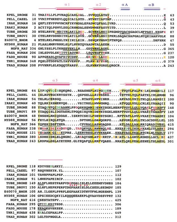
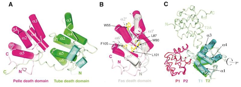
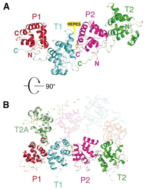
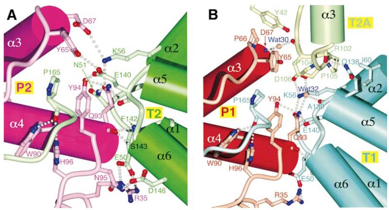
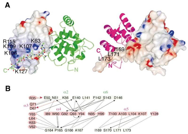
The interaction of the serine/threonine kinase Pelle and adaptor protein Tube through their N-terminal death domains leads to the nuclear translocation of the transcription factor Dorsal and activation of zygotic patterning genes during Drosophila embryogenesis. Crystal structure of the Pelle and Tube death domain heterodimer reveals that the two death domains adopt a six-helix bundle fold and are arranged in an open-ended linear array with plastic interfaces mediating their interactions. The Tube death domain has an insertion between helices 2 and 3, and a C-terminal tail making significant and indispensable contacts in the heterodimer. In vivo assays of Pelle and Tube mutants confirmed that the integrity of the major heterodimer interface is critical to the activity of these molecules.


Figures

Figure 1
Figure 1
Sequence Alignment of Death Domains Listed are death domains of D. melanogaster Pelle (sp|Q05652), C. elegans Pelle (gi|2814202), human IRAK (sp|P51617), IRAK-2 (gi|4504719), human D. melanogaster Tube (sp|P22812), D. virilis Tube (sp|Q08171), Bombyx mori Tube cDNA clone e40070 (gi|4156307), human MyD88 (gi|176309), rat P75 neurotrophin receptor (sp|P07174), human Fas (sp|P25445), mouse FADD (sp|Q61160), human TNF receptor 1 (sp|P19438), and human TRADD (sp|Q15628). Red cylinders indicate D. melanogaster Pelle death domain helices; blue cylinders indicate the two Tube death domain helix inserts. The helices of D. melanogaster Tube death domain, rat P75 receptor, human Fas, and mouse FADD are underlined. Highly conserved residues are highlighted in yellow, and conserved residues are highlighted in green. Residues are colored red, orange, or green where mutations cause complete loss, partial loss, or no obvious loss of function, respectively. The D. melanogaster Pelle or Tube residues at both P:T interfaces (see text) are indicated by red dots below the sequence; those at only one of the interfaces are indicated with orange dots.
Figure 2
Figure 2
The Six-Helix Bundle Fold of Pelle-DD and Tube-DD (A) P2:T2 dimer is shown with P2 in magenta and T2 in green. The inserted helices αA and αB of Tube-DD are colored cyan. (B) Superposition of Pelle-DD (magenta) and Fas-DD (gray). The view for Pelle-DD is the same as in (A). The five highly conserved residues are displayed as ball-and-stick models with those of Pelle-DD indicated by arrows. Only four residues, L87, W90, L101, and F105, are used to obtain this superposition. The helices of Fas-DD are marked. (C) P2:T2 dimer is superimposed onto P1:T1 using the Pelle-DD molecules of the respective dimers. P1, T1, P2, T2, and T2A are colored red, cyan, magenta, and green, respectively. The helices of T1 molecules are labeled. Figures prepared using Gl_render (Esser, 1999), BOBSCRIPT (Esnouf, 1997), MOLSCRIPT (Kraulis, 1991), and POV-ray (1998).
Figure 3
Figure 3
Interfaces between Pelle-DD and Tube-DD (A) The four death domains of the crystallographic asymmetric unit are colored according to Figure 2C. (B) The asymmetric unit is rotated 90° about the horizontal axis with an additional Tube (T2A) from another asymmetric unit (shown in low contrast) colored pale green.
Figure 4
Figure 4
Detailed View of the Dimer Interfaces (A) The P2:T2 dimer interface with interacting residues is displayed as a ball-and-stick model. Hydrogen bonds and salt bridges are indicated by gray dots. For clarity, part of the T2 C-terminal tail is omitted. The molecules are colored as in Figures 2C and 3. (B) The corresponding interface between P1 and T1 is shown with the intercalated T2A molecule. Part of the T1 C-terminal region is omitted for clarity.
Figure 5
Figure 5
The C-Terminal Tail of Tube-DD Contributes Significant Interaction Surface (A) The P2:T2 dimer: P2 is displayed as electrostatic surface (left) and magenta ribbon (right), T2 as green ribbon (left) and electrostatic surface (right), respectively. The color scale for the charge distribution extends from −10 kT/e (red) to +10 kT/e (blue) calculated using GRASP (Nicholls et al., 1991). The T2 carboxy-terminal hydrophobic residues in contact with P2, as well as charged residues of P2 surrounding the above region, are labeled on the respective surfaces. The T2 C-terminal tail is shown as a ball-and-stick model starting from residue 161 on the left. (B) The various contacts at the dimer interface are displayed as a schematic diagram with Pelle residues shaded magenta and Tube, green. Hydrogen bonds, salt bridges, and van der Waals interactions are indicated by black arrows (donor to acceptor), red arrows, and dotted lines, respectively.

Similar articles

Cited by

References

    1. Anderson KV, Nüsslein-Volhard C. Information for the dorsal-ventral pattern of the Drosophila embryo is stored as maternal mRNA. Nature. 1984;311:223–227. - PubMed
    1. Baker SJ, Reddy EP. Modulation of life and death by the TNF receptor superfamily. Oncogene. 1998;17:3261–3270. - PubMed
    1. Belvin MP, Jin Y, Anderson KV. Cactus protein degradation mediates Drosophila dorsal-ventral signaling. Gene Dev. 1995;9:783–793. - PubMed
    1. Bernstein FC, Koetzle TF, Williams JB, Meyer EF, Jr., Brice MD, Rodgers JR, Kennard O, Shimanouchi T, Tasumi M. The protein databank: a computer-based archival file for macromolecular structure. J. Mol. Biol. 1977;112:535–542. - PubMed
    1. Boldin MP, Mett IL, Varfolomeev EE, Chumakov I, Shemer-Avni Y, Camonis JH, Wallach D. Self-association of the “death domains” of the p55 tumor necrosis factor (TNF) receptor and Fas/APO1 prompts signaling for TNF and Fas/APO1 effects. J. Biol. Chem. 1995;270:387–391. - PubMed

Publication types

MeSH terms

Associated data