. 2000 Jan;74(1):57-64.
doi: 10.1128/jvi.74.1.57-64.2000.

Identifying the target cell in primary simian immunodeficiency virus (SIV) infection: highly activated memory CD4(+) T cells are rapidly eliminated in early SIV infection in vivo

Affiliations

Identifying the target cell in primary simian immunodeficiency virus (SIV) infection: highly activated memory CD4(+) T cells are rapidly eliminated in early SIV infection in vivo

R S Veazey et al. J Virol. 2000 Jan.

Abstract

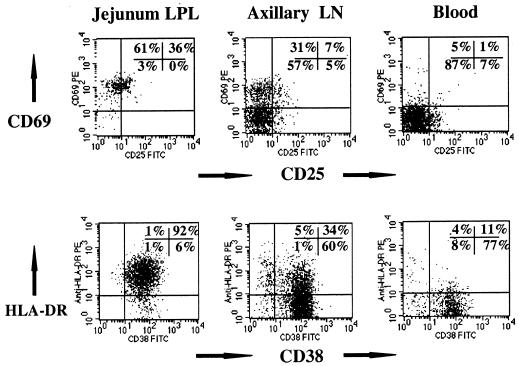
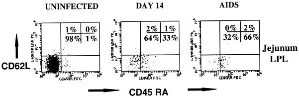
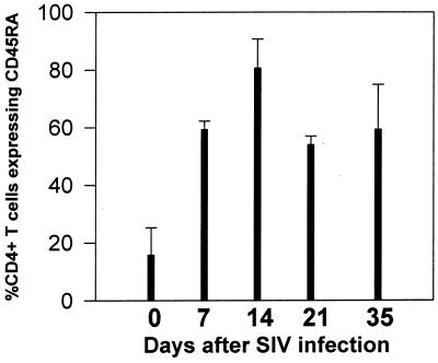
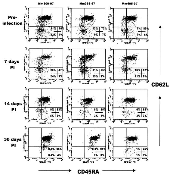
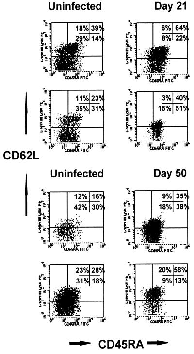
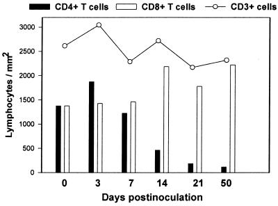
It has recently been shown that rapid and profound CD4(+) T-cell depletion occurs almost exclusively within the intestinal tract of simian immunodeficiency virus (SIV)-infected macaques within days of infection. Here we demonstrate (by three- and four-color flow cytometry) that this depletion is specific to a definable subset of CD4(+) T cells, namely, those having both a highly and/or acutely activated (CD69(+) CD38(+) HLA-DR(+)) and memory (CD45RA(-) Leu8(-)) phenotype. Moreover, we demonstrate that this subset of helper T cells is found primarily within the intestinal lamina propria. Viral tropism for this particular cell type (which has been previously suggested by various studies in vitro) could explain why profound CD4(+) T-cell depletion occurs in the intestine and not in peripheral lymphoid tissues in early SIV infection. Furthermore, we demonstrate that an acute loss of this specific subset of activated memory CD4(+) T cells may also be detected in peripheral blood and lymph nodes in early SIV infection. However, since this particular cell type is present in such small numbers in circulation, its loss does not significantly affect total CD4(+) T cell counts. This finding suggests that SIV and, presumably, human immunodeficiency virus specifically infect, replicate in, and eliminate definable subsets of CD4(+) T cells in vivo.


Figures

FIG. 1
FIG. 1
Comparison of lymphocyte activation (CD69, CD25, CD38, and HLA-DR expression) on CD4+ T cells from the intestinal lamina propria (left) to CD4+ T cells obtained from the axillary lymph node (LN; center) and blood (right) from an uninfected normal rhesus macaque. Note that essentially all intestinal CD4+ T cells are CD69+ HLA-DR+ CD38+. Plots were generated by gating first through lymphocytes and then through CD4+ cells.
FIG. 2
FIG. 2
Flow cytometry dot plots of CD45RA and L-selectin expression on intestinal lamina propria CD3+ CD4+ T cells from an uninfected macaque (left), a macaque infected for 14 days (center), and an animal with AIDS (right). Note that the vast majority of intestinal memory (CD45RA) cells are eliminated in early SIV infection, resulting in an increased proportion of naive (CD45RA+) cells remaining. Plots were generated by gating first through lymphocytes and then through CD3+ CD4+ DP T cells (four-color flow cytometry).
FIG. 3
FIG. 3
Sequential changes in CD45RA expression on duodenal CD4+ T cells in juvenile macaques infected with SIV. Intestinal biopsies were taken from the same three macaques before infection (day 0) and in the first few weeks after SIVmac251 infection. Bars represent the proportion of the total remaining CD4+ T cells that express CD45RA. Note that most intestinal CD4+ T cells are memory (CD45RA) cells before infection, whereas increased proportions of naive (CD45RA+) CD4+ T cells are detected in the intestine of the same animals within weeks of SIV infection. Each bar represents the mean of the three animals examined ± standard deviation.
FIG. 4
FIG. 4
Flow cytometry dot plots demonstrating a selective loss of memory CD4+ T cells in the peripheral blood of three macaques with early SIV infection. Each column shows the data from a single animal (animal numbers are listed above each column) examined before (top) and at 7, 14, and 30 days after SIV infection. Plots were generated by gating first through lymphocytes and then through CD4+ cells (three-color flow cytometry). Note that in each animal, a consistent loss of cells occurs in the upper left, lower left, and lower right quadrants by 14 days p.i., leaving only naive CD4+ (CD45RA+ CD62L+) T cells by 30 days p.i. (upper right quadrant). These results were representative of three separate experiments (n = 11).
FIG. 5
FIG. 5
Flow cytometry dot plots demonstrating a specific loss of memory CD4+ T cells in lymph nodes in early SIV infection. Plots on the left are from uninfected macaques, and those on the right are from different animals (cross-sectional study) infected with SIVmac239 for 21 or 50 days. Plots were generated by gating first through lymphocytes and then through CD3+ CD4+ cells (four-color flow cytometry). As in the blood, significantly fewer memory (CD45RA) cells are detected in lymph nodes from animals in early SIV infection than in those from uninfected animals. Note the decreased proportion of cells in the upper and lower left quadrants (memory cells) in the infected animals.
FIG. 6
FIG. 6
Estimation of the changes in the total numbers of T cells per square millimeter in the intestinal lamina propria in early SIV infection. Each time point represents the mean of two (infected) or four (uninfected control) animals. Morphometric analysis of CD3+ T cells in immunohistochemically stained jejunum sections was used to determine the numbers of CD3+ T cells per square millimeter of lamina propria (see text), and CD4+ and CD8+ T cells per square millimeter were determined by multiplying the number of CD3+ cells per square millimeter by the percentage of gated CD3+ lymphocytes coexpressing CD4 or CD8 in corresponding jejunum lamina propria as determined by flow cytometry (DP cells would be included in both bars). Note that a profound loss in CD4+ T cells per square millimeter occurs by 21 days after SIV infection, corresponding with an increase in absolute numbers of CD8+ T cells. Overall, this results in minimal changes in absolute numbers of CD3+ T cells in the lamina propria.

References

    1. Aiken C, Trono D. Nef stimulates human immunodeficiency virus type 1 proviral DNA synthesis. J Virol. 1995;69:5048–5056. - PMC - PubMed
    1. Akari H, Mori K, Otani I, Terao K, Ono F, Adachi A, Yoshikawa Y. Induction of MHC-IIDR expression on circulating CD8+ lymphocytes in macaques infected with SIVmac239 nef-open but not with its nef-deletion mutant. AIDS Res Hum Retroviruses. 1998;14:619–625. - PubMed
    1. Bell E B, Sparshott S M, Bunce C. CD4+ T-cell memory, CD45R subsets, and the persistence of antigen—a unifying concept. Immunol Today. 1998;19:60–64. - PubMed
    1. Berg M, Murakawa Y, Camerini D, James S P. Lamina propria lymphocytes are derived from circulating cells that lack the Leu-8 lymph node homing receptor. Gastroenterology. 1991;101:90–99. - PubMed
    1. Bjerke K, Brandtzaeg P, Fausa O. T cell distribution is different in follicle-associated epithelium of human Peyer's patches and villus epithelium. Clin Exp Immunol. 1988;74:270–275. - PMC - PubMed

Publication types

MeSH terms

Substances