. 2000 Jan;74(1):156-63.
doi: 10.1128/jvi.74.1.156-163.2000.

Ultrastructural and functional analyses of recombinant influenza virus ribonucleoproteins suggest dimerization of nucleoprotein during virus amplification

Affiliations

Ultrastructural and functional analyses of recombinant influenza virus ribonucleoproteins suggest dimerization of nucleoprotein during virus amplification

J Ortega et al. J Virol. 2000 Jan.

Abstract

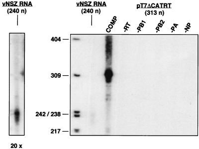
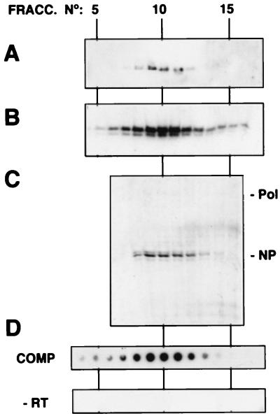
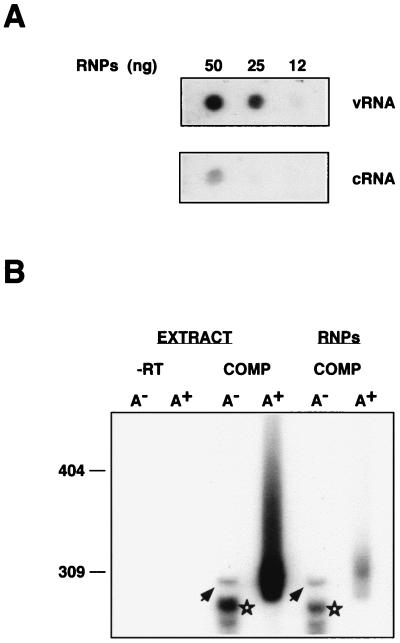
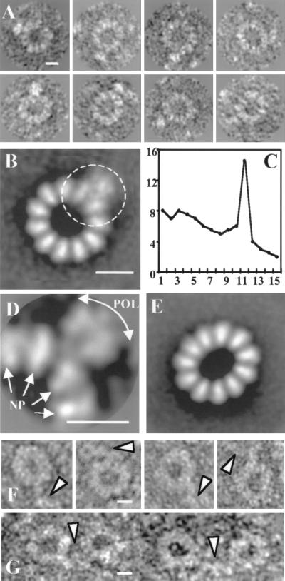
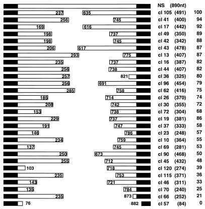
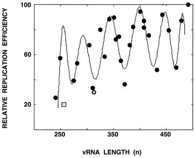
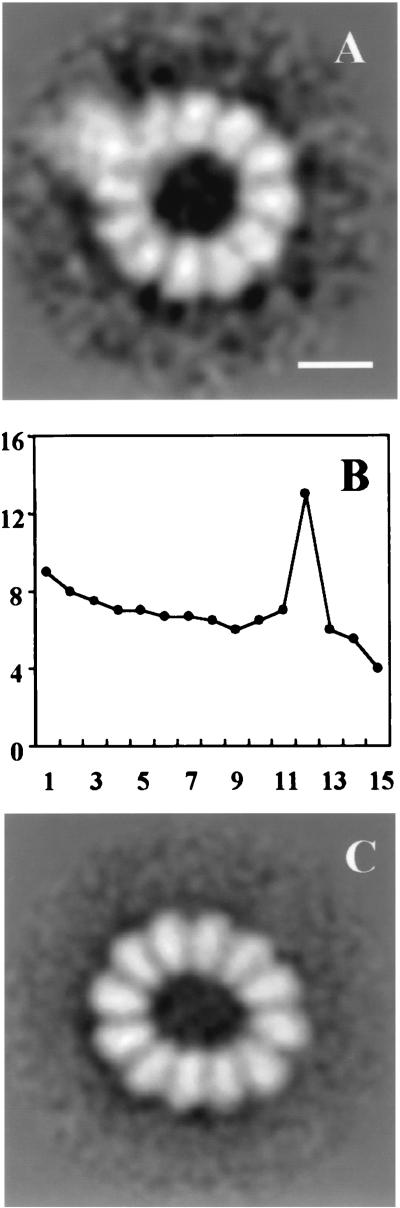
Influenza virus ribonucleoproteins (RNPs) were reconstituted in vivo from cloned cDNAs expressing the three polymerase subunits, the nucleoprotein (NP), and short template RNAs. The structure of purified RNPs was studied by electron microscopy and image processing. Circular and elliptic structures were obtained in which the NP and the polymerase complex could be defined. Comparison of the structure of RNPs of various lengths indicated that each NP monomer interacts with approximately 24 nucleotides. The analysis of the amplification of RNPs with different lengths showed that those with the highest replication efficiency contained an even number of NP monomers, suggesting that the NP is incorporated as dimers into newly synthesized RNPs.


Figures

FIG. 1
FIG. 1
Reconstitution of influenza virus mini-RNPs. Cultures of COS-1 cells were infected with vaccinia T7 virus and transfected with pGPB1, pGPB2, pGPA, and pGNPpolyA plasmids. The viral template was provided by simultaneous transfection of pT7NSΔCAT-RT plasmid or by delayed transfection of vNSZ RNA, as indicated in Materials and Methods. After incubation for 48 h, the viral RNPs were extracted and used for in vitro RNA synthesis by using ApG as primer. The RNA product was isolated and analyzed by electrophoresis on sequencing gels. The product obtained after transfection of pT7NSΔCAT-RT (313 nt) is shown (COMP), as well as the product generated by transfection of vNSZ RNA (240 nt) and those obtained when each of the elements of the systems was omitted. The panel to the left shows a 20× overexposure of the lane containing the vNSZ RNA product. The lengths of molecular weight markers are indicated to the left in nucleotides.
FIG. 2
FIG. 2
Purification of influenza virus mini-RNPs reconstituted in vivo. The viral RNPs reconstituted in vivo as indicated in Fig. 1 were purified by two successive glycerol gradients as indicated in Materials and Methods. The analyses corresponding to the fractionation of the second gradient are presented. Aliquots of each fraction were processed for Western blotting by using anti-PB1 (A) or anti-NP antibodies (B) or were analyzed by SDS-PAGE and Coomassie blue staining (C). The activity of each fraction was determined by in vitro transcription and TCA precipitation, filtration on a dot blot apparatus, and autoradiography (D, COMP). As a control, the activity of a parallel gradient with a sample in which no template was transfected (−RT) was determined. Pol and NP indicate the positions of the polymerase proteins and the NP in the gel, respectively.
FIG. 3
FIG. 3
Characterization of influenza virus RNPs reconstituted in vivo. Influenza virus RNPs were reconstituted and purified as indicated in Fig. 1 and 2. (A) The RNA contained in the purified RNPs was isolated and analyzed by dot blot hybridization by using positive (vRNA)- and negative (cRNA)-polarity riboprobes. (B) Either cellular extracts from infected and transfected cells (EXTRACT) or purified RNPs (RNPs) were used for in vitro transcription by using ApG as primer. The in vitro product was purified, fractionated on oligo(dT) cellulose into poly(A) (A) and poly(A)+ (A+) RNA, and analyzed on a sequencing gel. The products obtained when the complete reconstitution system was used are shown (COMP), as well as a control in which plasmid pT7NSΔCAT-RT was omitted (−RT). The arrowheads indicate the band corresponding to a full-length product. The stars show mRNA products deficient in polyadenylation. Numbers to the left refer to the mobility of molecular weight markers (in nucleotides).
FIG. 4
FIG. 4
Structure of influenza virus NSΔCAT RNP. Purified NSΔCAT RNPs were analyzed by electron microscopy after negative staining. The photographic plates were digitized, and 1,282 images from individual particles were stored and classified. Each homogeneous class was processed as described in Materials and Methods. (A) Gallery of circular RNPs. (B) Average image of the population of circular RNPs (average of 156 images; resolution, 30 Å). (C) Percentage of the total rotational power of the images presented in panel B plotted for the first 15 harmonics. (D) Average image with an alignment procedure that was centered on the polymerase complex (average of 55 images; resolution, 35 Å). (E) Average image of the population of ellipsoid RNPs (average of 871 images). (F) Gallery of images from RNP–anti-PB2 complexes. (G) Gallery of images from RNP–anti-PB2 complex dimers. Bar, 15 nm.
FIG. 5
FIG. 5
Relative amplification efficiency of influenza virus vRNPs reconstituted from templates of various lengths. Diagram of the structure of the NS segment (NS) and the collection of deletion derivatives, indicating the sequences deleted, the transcript length (in parentheses), and the relative activity in the in vitro transcription assay (taking clone 105 as a reference). The black regions at the ends represent the sequences conserved among all influenza virus RNA segments.
FIG. 6
FIG. 6
Length dependence of the efficiency of amplification of influenza virus RNPs. Graphic representation of the data presented in Fig. 5. The curve was adjusted to a polynomic function by using the program MATLAB 2.0.
FIG. 7
FIG. 7
Structure of clone 49 RNP. Purified clone 49 RNPs were analyzed by electron microscopy after negative staining. The photographic plates were digitized, and 386 images from individual particles were stored and classified. Each homogeneous class was processed as described in Materials and Methods. (A) Average image of the population of circular RNPs (average of 44 images; resolution, 35 Å). (B) Percentage of the total rotational power of the images presented in panel A plotted for the first 15 harmonics. (C) Average image of the population of ellipsoid RNPs (average of 336 images). Bar, 15 nm.

References

    1. Albo C, Valencia A, Portela A. Identification of an RNA binding region within the N-terminal third of the influenza A virus NP polypeptide. J Virol. 1995;69:3799–3806. - PMC - PubMed
    1. Ball L A. Cellular expression of a functional nodavirus RNA replicon from vaccinia virus vectors. J Virol. 1992;66:2335–2345. - PMC - PubMed
    1. Bárcena J, Ochoa M, de la Luna S, Melero J A, Nieto A, Ortín J, Portela A. Monoclonal antibodies against influenza virus PB2 and NP polypeptides interfere with the initiation step of viral mRNA synthesis in vitro. J Virol. 1994;68:6900–6909. - PMC - PubMed
    1. Baudin F, Bach C, Cusack S, Ruigrok R W. Structure of influenza virus RNP. I. Influenza virus nucleoprotein melts secondary structure in panhandle RNA and exposes the bases to the solvent. EMBO J. 1994;13:3158–3165. - PMC - PubMed
    1. Biswas S K, Nayak D P. Mutational analysis of the conserved motifs of influenza A virus polymerase basic protein 1. J Virol. 1994;68:1819–1826. - PMC - PubMed

Publication types