. 2000 Jan;74(1):164-72.
doi: 10.1128/jvi.74.1.164-172.2000.

RNA dimerization defect in a Rous sarcoma virus matrix mutant

Affiliations

RNA dimerization defect in a Rous sarcoma virus matrix mutant

L J Parent et al. J Virol. 2000 Jan.

Abstract

The retrovirus matrix (MA) sequence of the Gag polyprotein has been shown to contain functions required for membrane targeting and binding during particle assembly and budding. Additional functions for MA have been proposed based on the existence of MA mutants in Rous sarcoma virus (RSV), murine leukemia virus, human immunodeficiency virus type 1, and human T-cell leukemia virus type 1 that lack infectivity even though they release particles of normal composition. Here we describe an RSV MA mutant with a surprising and previously unreported phenotype. In the mutant known as Myr1E, the small membrane-binding domain of the Src oncoprotein has been added as an N-terminal extension of Gag. While Myr1E is not infectious, full infectivity can be reestablished by a single amino acid substitution in the Src sequence (G2E), which eliminates the addition of myristic acid and the membrane-binding capacity of this foreign sequence. The presence of myristic acid at the N terminus of the Myr1E Gag protein does not explain its replication defect, because other myristylated derivatives of RSV Gag are fully infectious (e.g., Myr2 [C. R. Erdie and J. W. Wills, J. Virol. 64:5204-5208, 1990]). Biochemical analyses of Myr1E particles reveal that they contain wild-type levels of the Gag cleavage products, Env glycoproteins, and reverse transcriptase activity when measured on an exogenous template. Genomic RNA incorporation appears to be mildly reduced compared to the wild-type level. Unexpectedly, RNA isolated from Myr1E particles is monomeric when analyzed on nondenaturing Northern blots. Importantly, the insertional mutation does not lie within previously identified dimer linkage sites. In spite of the dimerization defect, the genomic RNA from Myr1E particles serves efficiently as a template for reverse transcription as measured by an endogenous reverse transcriptase assay. In marked contrast, after infection of avian cells, the products of reverse transcription are nearly undetectable. These findings might be explained either by the loss of a normal function of MA needed in the formation or stabilization of RNA dimers or by the interference in such events by the mutant MA molecules. It is possible that Myr1E viruses package a single copy of viral RNA.


Figures

FIG. 1
FIG. 1
Schematic representation of wild-type and mutant MA proteins. The N-terminal region of the RSV Gag protein is shown at the top with the MA, p2, and p10 cleavage products indicated; the numbers below refer to amino acid residues. The M domain, as indicated, consists of the first 85 residues of MA. Expanded below are the N-terminal amino acid sequences of wild-type (WT) and mutant proteins. Residues identical to WT are indicated by dots. Boxes are drawn around residues derived from the Src membrane-binding domain, with the squiggle representing myristic acid modification. Myr2 has an E2G substitution, resulting in myristylation and removal of initiator methionine. In Myr1, the first 10 gag codons were replaced with the sequence encoding the myristylated Src N terminus. Myr1− has the wild-type glutamate substituted at position 2 of Myr1, resulting in the prevention of myristylation and the retention of methionine. Myr1E has nine codons specifying the Src M domain inserted after the gag AUG so that the Src sequence is extended from the Gag N terminus. In Myr1E−, myristylation is abolished by a G2E substitution. The ability of each virus to bud and to infect avian cells is indicated by a plus sign (equivalent to wild-type level), minus sign (≤1% of wild-type level), or NT (not tested).
FIG. 2
FIG. 2
Effects of MA alterations on particle assembly and myristylation. (A) Analysis of budding in COS-1 cells. Transfected COS-1 cells were radiolabeled with [35S]methionine for 2.5 h, lysates and media were separated, and viral proteins were detected by immunoprecipitation with anti-RSV serum. Positions of the bands corresponding to Gag precursor, CA, MA, and PR proteins are indicated at the left. untr'f, untransfected. (B) Myristate labeling. Duplicate plates of COS-1 cells were transfected, labeled for 70 min with [35S]methionine or [3H]myristic acid, and immunoprecipitated as for panel A. (C) Budding in avian cells. QT6 cells were transfected with wild-type (WT) and mutant proviral plasmids and analyzed as for panel A. The position of the Gag-Pol protein is indicated at the left, and positions of molecular weight standards are shown at the right.
FIG. 3
FIG. 3
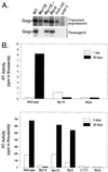
Persistence of virus production in long-term cultures. (A) Duplicate plates of QT6 cells were transfected with proviral constructs expressing the indicated gag alleles. At 18 h posttransfection, the Gag precursor in cell lysates was detected after a 1-h labeling period by immunoprecipitation with RSV antiserum (transient expression). Persistent expression of Gag was measured similarly after passage 6. non-inf., noninfectious CA mutant L171I, included as a negative control; untr'f, untransfected. (B) In two separate experiments, infections of QT6 cells were initiated by transfection as for panel A; then the accumulation of RT activity in the culture medium was monitored over a period of 30 or 33 days posttransfection. The large difference between the two experiments in the scale on the RT activity axis was due to differences in specific activity of the isotope and in the particle concentration factor.
FIG. 4
FIG. 4
Biochemical properties of virus particles. (A) Envelope incorporation. Equivalent amounts of virions from culture supernatants were collected by ultracentrifugation from QT6 cells transfected with mutant or wild-type (WT) proviral plasmids. Levels of Env (SU) expression were detected by immunoblotting with anti-Env serum. untr'f, untransfected. (B) Gag processing. Virus particles from panel A were analyzed by immunoblotting with anti-RSV serum to detect processed Gag proteins CA and MA. (C) RNA genome incorporation. Virus particles were normalized for RT content, RNA was purified, serial fourfold dilutions were performed, and the RNA was applied to a slot blot apparatus, bound to nitrocellulose, and detected with a radiolabled gag riboprobe.
FIG. 5
FIG. 5
Detection of viral DNA after infection. (A) Virus particles produced from stably expressing cell lines were normalized, concentrated, and used to infect QT6 cells. Low-molecular-weight DNA was isolated and used as a template in PCRs with primers specific for minus-strand strong-stop DNA as described in Materials and Methods. In both panels, molecular size markers were run in the right lane (ladder). The PCR product amplified from minus-strand strong-stop DNA is predicted to be 86 bp in length with our primers. cl., clone. (B) Quantitative analysis of strong-stop DNA. Low-molecular-weight DNA isolated as described for panel A was serially diluted as indicated and amplified as described above. Virus particles used for infection were derived from independently derived cell lines (clones 1 and 2) stably expressing the myr1e genome.
FIG. 6
FIG. 6
ERT activity. Permeabilized visions were incubated with nucleotides, and viral DNA was isolated and subjected to PCR analysis as for Fig. 5. (A) Intravirion minus-strand strong-stop DNA for particles isolated from stable cell lines. Molecular size markers were run in the right lane (ladder). (B) Serial 10-fold dilutions of ERT products to amplify sequences corresponding to minus-strand strong-stop (86-bp), first-strand switch (117-bp), and second-strand switch (300-bp) DNA products.
FIG. 7
FIG. 7
Nondenaturing Northern blot analysis of viral RNA. Genomic RNA was isolated from virus particles produced by transient transfection of proviral plasmids (A) or from two independently arising stably producing cell lines (clones [cl.] 1 and 2) (B). The positions of dimers (D) and monomers (M) are indicated by arrows.

Similar articles

Cited by

References

    1. Aronoff R, Hajjar A M, Linial M L. Avian retroviral encapsidation: reexamination of functional 5′ RNA sequences and the role of the nucleocapsid Cys-His motifs. J Virol. 1993;67:178–188. - PMC - PubMed
    1. Aronoff R, Linial M L. Specificity of retroviral packaging. J Virol. 1991;65:71–80. - PMC - PubMed
    1. Bennett R B, Wills J W. Conditions for copackaging Rous sarcoma virus and murine leukemia virus Gag proteins during retroviral budding. J Virol. 1999;73:2045–2051. - PMC - PubMed
    1. Bennett R P, Nelle T D, Wills J W. Functional chimeras of the Rous sarcoma virus and human immunodeficiency virus Gag proteins. J Virol. 1993;67:6487–6498. - PMC - PubMed
    1. Berkowitz R, Fisher J, Goff S P. RNA packaging. In: Krausslich H-G, editor. Morphogenesis and maturation of retroviruses. Berlin, Germany: Springer-Verlag; 1996. pp. 177–218.

Publication types

LinkOut - more resources