Immunohistochemical analysis of primary sensory neurons latently infected with herpes simplex virus type 1
- PMID: 10590108
- PMCID: PMC111530
- DOI: 10.1128/jvi.74.1.209-217.2000
Immunohistochemical analysis of primary sensory neurons latently infected with herpes simplex virus type 1
Abstract
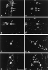
We characterized the populations of primary sensory neurons that become latently infected with herpes simplex virus (HSV) following peripheral inoculation. Twenty-one days after ocular inoculation with HSV strain KOS, 81% of latency-associated transcript (LAT)-positive trigeminal ganglion (TG) neurons coexpressed SSEA3, 71% coexpressed Trk(A) (the high-affinity nerve growth factor receptor), and 68% coexpressed antigen recognized by monoclonal antibody (MAb) A5; less than 5% coexpressed antigen recognized by MAb KH10. The distribution of LAT-positive, latently infected TG neurons contrasted sharply with (i) the overall distribution of neuronal phenotypes in latently infected TG and (ii) the neuronal distribution of viral antigen in productively infected TG. Similar results were obtained following ocular and footpad inoculation with KOS/62, a LAT deletion mutant in which the LAT promoter is used to drive expression of the Escherichia coli lacZ gene. Thus, although all neuronal populations within primary sensory ganglia appear to be capable of supporting a productive infection with HSV, some neuronal phenotypes are more permissive for establishment of a latent infection with LAT expression than others. Furthermore, expression of HSV LAT does not appear to play a role in this process. These findings indicate that there are marked differences in the outcome of HSV infection among the different neuronal populations in the TG and highlight the key role that the host neuron may play in regulating the repertoire of viral gene expression during the establishment of HSV latent infection.
Figures


References
-
- Clements G B, Stow N D. A herpes simplex virus type 1 mutant containing a deletion within immediate early gene 1 is latency-competent in mice. J Gen Virol. 1989;70:2501–2506. - PubMed
-
- Cunningham E T, Jr, DeSouza E B. Localization of type I IL-1 receptor mRNA in brain and endocrine tissues using in situ hybridization histochemistry. In: DeSouza E B, editor. Methods in neuroscience, part A. Neurobiology of cytokines. New York, N.Y: CRC Press; 1994. pp. 112–127.
Publication types
MeSH terms
Grants and funding
LinkOut - more resources
Full Text Sources
Research Materials
Miscellaneous

