. 2000 Jan;74(1):218-27.
doi: 10.1128/jvi.74.1.218-227.2000.

Initial events in infectious salmon anemia virus infection: evidence for the requirement of a low-pH step

Affiliations

Initial events in infectious salmon anemia virus infection: evidence for the requirement of a low-pH step

T M Eliassen et al. J Virol. 2000 Jan.

Abstract

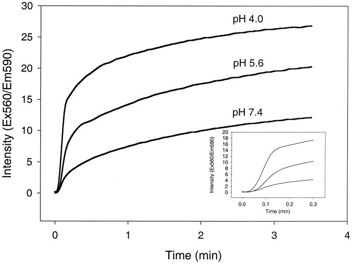
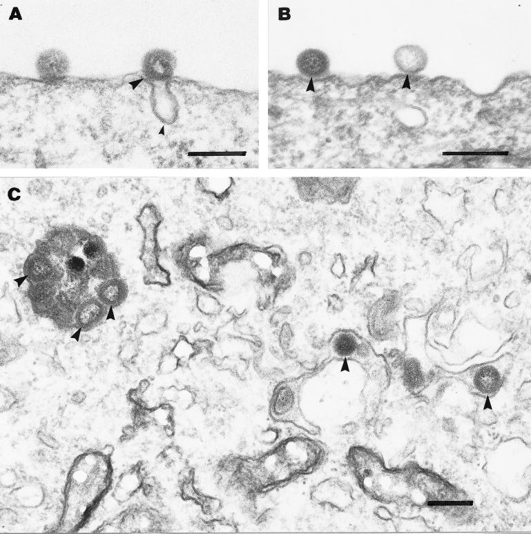
We have investigated the initial steps in the interaction between infectious salmon anemia virus (ISAV) and cultured cells from Atlantic salmon (SHK-1 cell line). Using radioactively or fluorescently labelled viral particles we have studied the binding and fusion kinetics and the effect of pH on binding, uptake, and fusion of ISAV to SHK-1 cells and liposomes. As pH in the medium was reduced from 7.5 to 4.5, the association of virus to the cells was nearly doubled. The same effect of pH was observed when fusion between ISAV and liposomes was analyzed. In addition, the binding of ISAV to intact SHK-1 cells and to cell membrane proteins blotted onto filters was neuraminidase sensitive. However, the increased binding induced by low pH was not neuraminidase sensitive, probably reflecting activation of a fusion peptide at low pH. By using confocal fluorescence microscopy, the increased fusion of fluorescently labelled ISAV with the plasma membrane due to low pH could be demonstrated. When vacuolar pH in the cells was raised during inoculation with chloroquine or ammonium chloride, both electron and confocal microscopy showed accumulation of ISAV in endosomes and lysosomes. Production of infectious virus could be increased by lowering the extracellular pH during infection. Furthermore, chloroquine present during virus inoculation also caused a reduction in the synthesis of viral proteins in ISAV-infected cells as well as in the production of infective virus. These results indicate that ISAV binds to sialic acid residues on the cell surface and that the fusion between virus and cell membrane takes place in the acid environment of endosomes. This provides further evidence for a high degree of similarity between ISAV and influenza virus and extends the basis for the classification of this virus as a member of the Orthomyxoviridae family.


Figures

FIG. 1
FIG. 1
Interaction of ISAV with SHK-1 cells. (A) Kinetics of ISAV binding to SHK-1 cells at pH 7.4. The cells were incubated with trace quantities of 125I-labelled virus at 0°C. Cell-associated radioactivity was determined at the indicated times. Shown is a typical experiment (wells in triplicate). (B) Effect of pH on ISAV binding to SHK-1 cells at 0°C. The cells were incubated with 125I-labelled virus for 3 h at 0°C in L-15 medium adjusted to the indicated pH. After 3 h the cells were washed three times with binding medium and solubilized in 0.1 N NaOH–1% SDS and cell-associated virus radioactivity was determined. Data are expressed as percentages of control binding at pH 7.4 in each experiment. Values are the means of three different experiments +/− standard deviations (SD). (C) Effect of pH on neuraminidase-sensitive binding of ISAV to SHK-1 cells. Trace quantities of 125I-labelled ISAV were allowed to bind to SHK-1 cells for 3 h at 0°C and pH 4.5 or 7.4. After being washed, cells were treated with neuraminidase (5 mg/ml) in L-15 medium for 90 min at 0°C. The cells were washed twice with L-15 medium before cell-associated radioactivity was determined. Data are expressed as percentages of control. Values are the means of three different experiments +/− SD.
FIG. 2
FIG. 2
Binding of 125I-labelled ISAV to membrane proteins. (A) Membrane fractions (50 μg of protein/lane) from SHK-1 cells (lanes 1 and 2), salmon liver (lanes 3 and 4), and rainbow trout liver (lanes 5 and 6) were separated by SDS-PAGE, electroblotted, blocked with 5% bovine serum albumin in PBS containing Tween 80 for 1 h, and incubated overnight with 125I-labelled virus. Binding was detected with autoradiographic film for 24 h. (B) Membrane fractions (50 μg/lane) from salmon liver electrophoresed and blotted with rab5 antibody before (control) or after neuraminidase treatment (5 mg/ml in L15 medium for 90 min) of the filter. (C) Mucin (100 μg), dissolved in 10 μl of SDS sample buffer was applied to a nitrocellulose filter and blocked for 1 h with 5% dry milk. The filter was then incubated with trace amounts of 125I-labelled ISAV overnight. After a wash, binding was detected by autoradiography.
FIG. 3
FIG. 3
Fusion of fluorescent ISAV to SHK-1 cells. SHK-1 cells were grown on glass coverslips and incubated with R18-labelled ISAV for 4 h at pH 7.5 (A) or 4.5 (B) or in the presence of 0.1 mM chloroquine (C) or 0.1 mM ammonium chloride (D). The cells were washed and fixed with 2% paraformaldehyde in PBS for 10 min and photographed with a Leica confocal fluorescence microscope.
FIG. 4
FIG. 4
Fusion of R18-labelled ISAV with liposomes. Liposomes were diluted to a lipid concentration of 0.3 mM in the three different buffers mentioned in “Preparation of liposomes.” The solution (90 μl) was added to a cuvette containing 10 μl (1.4 mg of viral protein/ml) of R18-labelled ISAV. The increase in rhodamine fluorescence was measured with a Perkin-Elmer LS50B luminescence spectrometer (excitation [Ex], 560 nm; emission [Em], 590 nm). Each sample was assayed three times, and the average intensity values were plotted against time.
FIG. 5
FIG. 5
Electron micrographs of the early steps in ISAV infection. (A and B) Association of ISAV (big arrowheads in all panels) with the plasma membrane after 4 h of binding at 4°C. Virus was found associated with either invaginations presumably representing caveolae (small arrowhead) (A) or flat plasma membrane stretches without any visible coating (B). Further incubation of cells for 4 h with chloroquine (0.1 mM) at 15°C led to intracellular accumulation of ISAV in vesicular and tubular structures (C). Bars, 200 nm.
FIG. 6
FIG. 6
Effect of pH on fusion of ISAV with the plasma membrane. ISAV was bound to SHK-1 cells for 4 h at 0°C in BM (pH 7.4). The cells were then washed and incubated with biotin anti-ISAV MAb (diluted 1:200 in serum-free L-15 medium) for 1 h at 0°C. After being washed, the samples were incubated with streptavidin-conjugated colloidal gold (10 nm) (diluted 1:50 in serum-free L-15 medium) for 1 h at 0°C. The cells were then chased for 2 h before fixation. (A) Association of ISAV with an invagination of the plasma membrane of SHK-1 cells. (B) Internalized ISAV in a vesicular structure within an SHK-1 cell. (C) Samples exposed to acidic binding medium (pH 4.5) for 10 min after binding and immunolabelling. The fusion of ISAV with the plasma membrane is shown. In some cases, clear continuity between the cell and virus membrane was observed. The virus particles also seemed to aggregate and fuse with each other at the plasma membrane. Bars, 200 nm.
FIG. 7
FIG. 7
ISAV entry from endosomes and lysosomes. ISAV was bound to SHK-1 cells for 4 h at 0°C in BM (pH 7.4). After being washed the cells were chased for 2 h at 15°C before fixation. Shown is internalized virus fusing with the vesicular membrane. Clear continuity between the virus membrane and the vesicular membrane was observed. Bar, 200 nm.
FIG. 8
FIG. 8
Effect of pH perturbants on ISAV production in SHK-1 cells. (A) Chloroquine (10 or 100 μM) was present during inoculation (4 h) or after inoculation with virus as detailed in Materials and Methods. The infectious virus titer of the medium was determined at the indicated times after infection. TCID50, 50% tissue culture infective dose. (B) Comparison of the effect of chloroquine (100 μM) or bafilomycin A1 (1 μM) on ISAV production in SHK-1 cells. The inhibitors were present during (4 h) or after inoculation (4 h). The infectious titer of the medium was determined at day 2 p.i.
FIG. 9
FIG. 9
Effect of chloroquine on synthesis of viral polypeptides in SHK-1 cells. The polypeptides were analyzed by SDS-PAGE and autoradiography on day 4 p.i. following a 24-h incubation with [35S]methionine. Lanes 1 to 3, ISAV-infected cells; lanes 4 to 6, noninfected cells. Lanes 1 and 4, no chloroquine; lanes 2 and 5, chloroquine (100 μM) present during inoculation (4 h); lanes 3 and 6, chloroquine (100 μM) present after inoculation (4 h). Lines at the left indicate positions of molecular mass markers (70 [upper] and 25 kDa [lower]).

References

    1. Bagai S, Lamb R A. Quantitative measurement of paramyxovirus fusion: differences in requirements of glycoproteins between simian virus 5 and human parainfluenza virus 3 or Newcastle disease virus. J Virol. 1995;69:6712–6719. - PMC - PubMed
    1. Berg T, Tolleshaug H. The effects of ammonium ions and chloroquine on uptake and degradation of 125I-labeled asialo-fetuin in isolated rat hepatocytes. Biochem Pharmacol. 1980;29:917–925. - PubMed
    1. Blumenthal R, Bali-Puri A, Walter A, Covell D, Eidelman O. pH-dependent fusion of vesicular stomatitis virus with Vero cells. Measurement by dequenching of octadecyl rhodamine fluorescence. J Biol Chem. 1987;262:13614–13619. . (Erratum, 263:588, 1988.) - PubMed
    1. Bullough P A, Hughson F M, Skehel J J, Wiley D C. Structure of influenza haemagglutinin at the pH of membrane fusion. Nature. 1994;371:37–43. - PubMed
    1. Crider B P, Xie X S, Stone D K. Bafilomycin inhibits proton flow through the H+ channel of vacuolar proton pumps. J Biol Chem. 1994;269:17379–17381. - PubMed

Publication types

MeSH terms