. 2000 Jan;74(1):272-80.
doi: 10.1128/jvi.74.1.272-280.2000.

Foot-and-mouth disease virus 3C protease induces cleavage of translation initiation factors eIF4A and eIF4G within infected cells

Affiliations

Foot-and-mouth disease virus 3C protease induces cleavage of translation initiation factors eIF4A and eIF4G within infected cells

G J Belsham et al. J Virol. 2000 Jan.

Abstract

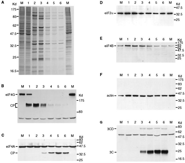
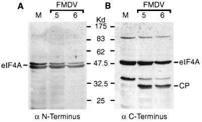
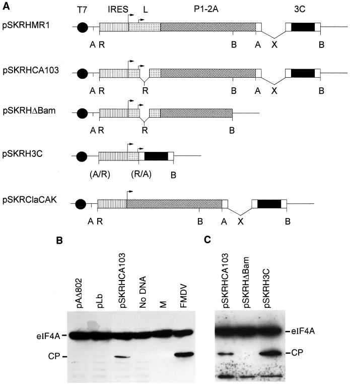
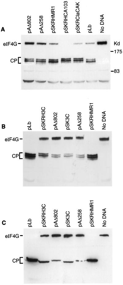
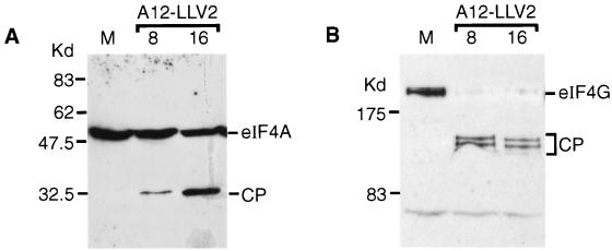
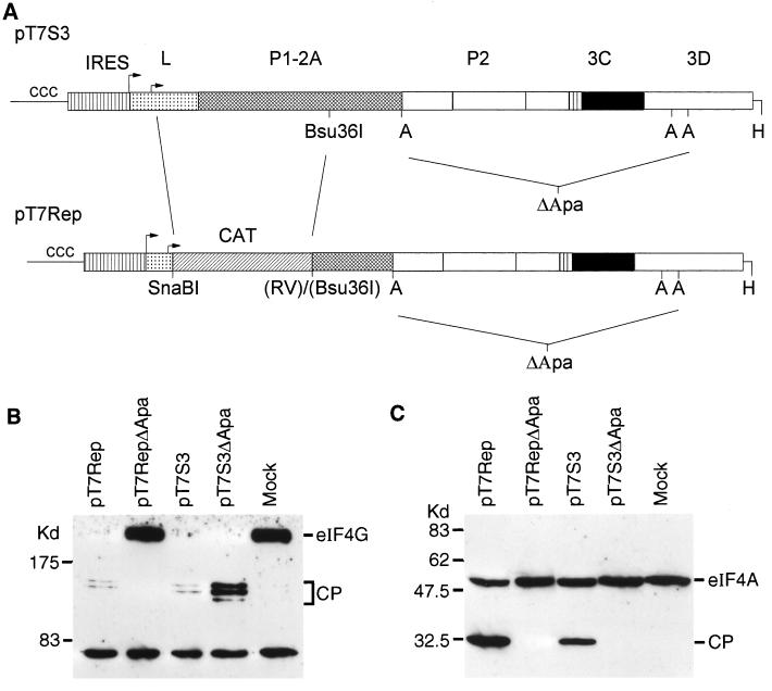
Infection of cells by foot-and-mouth disease virus (FMDV) results in the rapid inhibition of host cell protein synthesis. This process is accompanied by the early cleavage of the translation initiation factor eIF4G, a component of the cap-binding complex eIF4F. This cleavage is mediated by the leader (L) protease. Subsequently, as the virus proteins accumulate, secondary cleavages of eIF4G occur. Furthermore, eIF4A (46 kDa), a second component of eIF4F, is also cleaved in these later stages of the infection cycle. The 33-kDa cleavage product of eIF4A has lost a fragment from its N terminus. Transient-expression assays demonstrated that eIF4A was not cleaved in the presence of FMDV L or with the poliovirus 2A protease (which also mediates eIF4G cleavage) but was cleaved when the FMDV 3C protease was expressed. The FMDV 3C protease was also shown in such assays to induce cleavage of eIF4G, resulting in the production of cleavage products different from those generated by the L protease. Consistent with these results, within cells infected with a mutant FMDV lacking the L protease or within cells containing an FMDV replicon lacking L-P1 coding sequences it was again shown that eIF4A and eIF4G were cleaved.


Figures

FIG. 1
FIG. 1
Analysis of translation initiation factors within FMDV-infected cells. BHK cells were infected with FMDV. (A) At the indicated times (hours postinfection), cells were transferred to [35S]EXPRESS-containing medium and incubation was continued for a further 15 min. Cell extracts were prepared and analyzed by SDS-PAGE (10% polyacrylamide). (B to G) Alternatively, cells infected in parallel with those analyzed in panel A were harvested at the same times without metabolic labelling. Cell extracts were analyzed by SDS-PAGE (6% [B] or 10% [C to G] polyacrylamide) and immunoblotting with antibodies specific for the N-terminal region of eIF4G (B), eIF4A (C), eIF2α (D), eIF4B (E), actin (F), or FMDV 3C (G). Mock-infected BHK cell extracts (lanes M) were analyzed in parallel in each case.
FIG. 2
FIG. 2
Characterization of eIF4A cleavage product in FMDV-infected cells. BHK cell extracts from mock-infected cells (lane M) or FMDV-infected cells (5 or 6 h postinfection as indicated) were analyzed by SDS-PAGE and immunoblotting with rabbit antisera specific for the N-terminal region peptide SQDSRSDNGPDGMEPEK (A) or the C-terminal region peptide DLPANRENYIHRTGRGGRFGRK (B) of the human eIF4AI sequence (kindly provided by S. J. Morley, University of Sussex). Detection on X-ray film was achieved by using peroxidase-labelled anti-rabbit IgG antibody and chemiluminescence reagents.
FIG. 3
FIG. 3
Identification of eIF4A-directed protease. (A) Structures of plasmids expressing FMDV cDNA cassettes encoding the indicated proteases. All plasmids contain the T7 promoter and the FMDV IRES element. Restriction enzyme sites: A, ApaI; R, EcoRI; B, BamHI; X, XhoI. Parentheses indicate that the site is lost. Note that plasmids pSKRHMR1 and pSKRHCA103 were reported previously (3) and are included for completeness. (B) BHK cells were infected with vTF7-3 (22) and transfected with the indicated plasmids. After 20 h, cell extracts were prepared and analyzed by SDS-PAGE (10% polyacrylamide) and immunoblotting with anti-eIF4A. Samples from mock (lane M)- or FMDV-infected BHK cells were analyzed on the same gel. (C) vTF7-3-infected HTK-143 cells were transfected with the indicated plasmids (see panel A) and analyzed as for panel B.
FIG. 4
FIG. 4
FMDV 3C induces cleavage of eIF4G (A). BHK cells were infected with vTF7-3 (as for Fig. 3) and transfected with the indicated plasmids (Fig. 3A). After 20 h, cell extracts were prepared and analyzed by SDS-PAGE (6% polyacrylamide) and immunoblotting with an N-terminal region-specific anti-eIF4G antiserum. (B and C) In a similar experiment vTF7-3-infected BHK cells were transfected with the indicated plasmids, and cell extracts were analyzed by SDS-PAGE (6% polyacrylamide) and immunoblotting with antisera specific for the N-terminal region of eIF4G (B) or the C-terminal fragment of eIF4G (C).
FIG. 5
FIG. 5
eIF4A and eIF4G cleavage in A12-LLV2 FMDV-infected cells. Mock (lanes M)- or A12-LLV2 FMDV-infected BHK cells were harvested at 8 or 16 h postinfection as indicated. Cell extracts were prepared and analyzed by SDS-PAGE and immunoblotting with anti-eIF4A (A) or anti-eIF4G (B) as for Fig. 3 and 4.
FIG. 6
FIG. 6
L-deficient FMDV replicon induces specific eIF4A and eIF4G cleavage. (A) The structure of the FMDV replicon (pT7Rep) is indicated together with the parental infectious cDNA pT7S3 (18) and the sequences removed (by digestion of the cDNA with ApaI and religation) to generate the nonreplicating derivatives of the replicon (pT7RepΔApa) and its parent (pT7S3ΔApa). Restriction sites: A, ApaI; H, HpaI; RV, EcoRV. (B and C) RNA transcripts prepared from HpaI-linearized plasmids by using T7 RNA polymerase were electroporated into BHK cells. After 6 h cell extracts were prepared and analyzed for eIF4G (B) and eIF4A (C) as for Fig. 3 and 4.

References

    1. Belsham G J. Dual initiation sites of protein synthesis on foot-and-mouth disease virus RNA are selected following internal entry and scanning of ribosomes in vivo. EMBO J. 1992;11:1105–1110. - PMC - PubMed
    1. Belsham G J. Distinctive features of foot-and-mouth disease virus, a member of the picornavirus family; aspects of virus protein synthesis, protein processing and structure. Prog Biophys Mol Biol. 1993;60:241–260. - PMC - PubMed
    1. Belsham G J, Brangwyn J K. A region of the 5′ noncoding region of foot-and-mouth disease virus RNA directs efficient internal initiation of protein synthesis within cells: involvement with the role of L protease in translational control. J Virol. 1990;64:5389–5395. - PMC - PubMed
    1. Belsham G J, Abrams C A, King A M Q, Roosien J, Vlak J M. Myristoylation of foot-and-mouth disease virus capsid protein precursors is independent of other viral proteins and occurs in both mammalian and insect cells. J Gen Virol. 1991;72:747–751. - PubMed
    1. Belsham G J. Analysis of picornavirus internal ribosome entry site function in vivo. In: Richter J, editor. mRNA formation and function. New York, N.Y: Academic Press; 1997. pp. 323–340.

Publication types

LinkOut - more resources