. 2000 Jan;74(1):281-94.
doi: 10.1128/jvi.74.1.281-294.2000.

Conditional site-specific integration into human chromosome 19 by using a ligand-dependent chimeric adeno-associated virus/Rep protein

Affiliations

Conditional site-specific integration into human chromosome 19 by using a ligand-dependent chimeric adeno-associated virus/Rep protein

D Rinaudo et al. J Virol. 2000 Jan.

Abstract

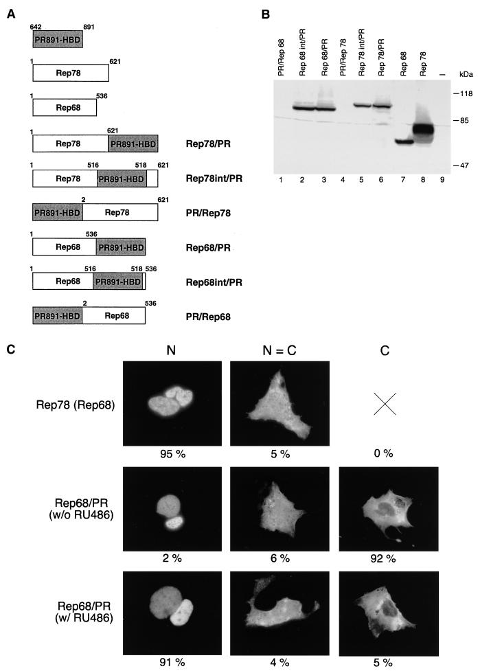
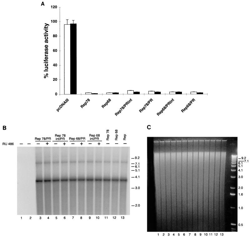
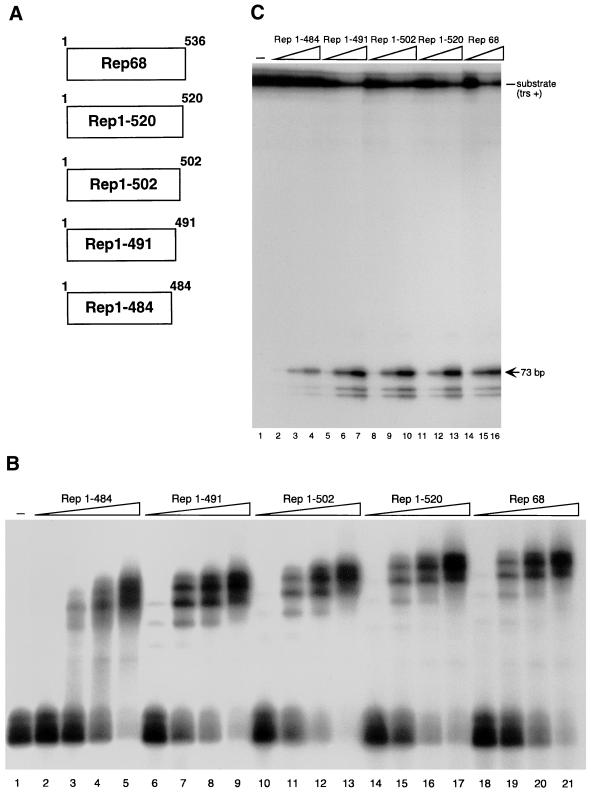
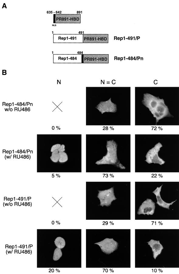
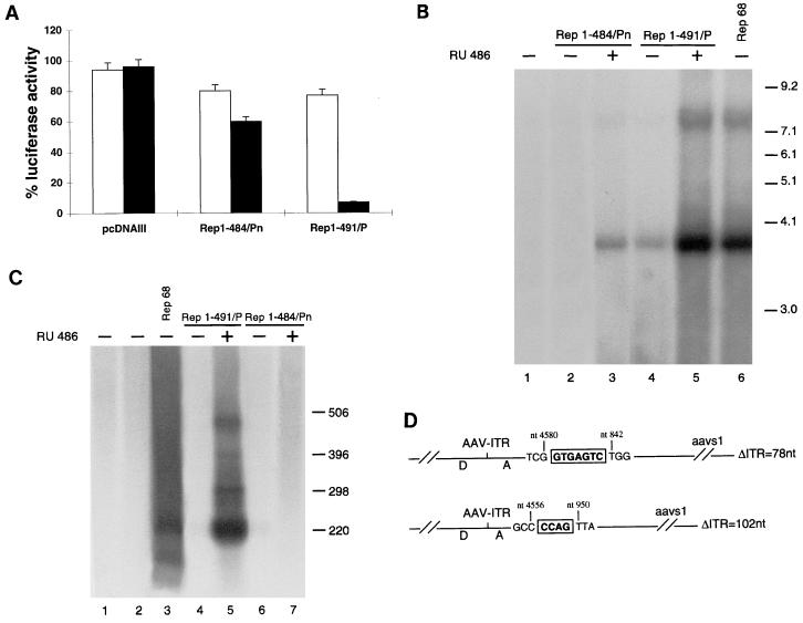
It is of great interest for gene therapy to develop vectors that drive the insertion of a therapeutic gene into a chosen specific site on the cellular genome. Adeno-associated virus (AAV) is unique among mammalian viruses in that it integrates into a distinct region of human chromosome 19 (integration site AAVS1). The inverted terminal repeats (ITRs) flanking the AAV genome and the AAV-encoded nonstructural proteins Rep78 and/or Rep68 are the only viral elements necessary and sufficient for site-specific integration. However, it is also known that unrestrained Rep activity may cause nonspecific genomic rearrangements at AAVS1 and/or have detrimental effects on cell physiology. In this paper we describe the generation of a ligand-dependent form of Rep, obtained by fusing a C-terminally deleted Rep68 with a truncated form of the hormone binding domain of the human progesterone receptor, which does not bind progesterone but binds only its synthetic antagonist RU486. The activity of this chimeric protein, named Rep1-491/P, is highly dependent on RU486 in various assays: in particular, it triggers site-specific integration at AAVS1 of an ITR-flanked cassette in a ligand-dependent manner, as efficiently as wild-type Rep68 but without generating unwanted genomic rearrangement at AAVS1.


Figures

FIG. 1
FIG. 1
Structure, expression, and intracellular distribution of fusion constructs derived from wt Rep78 and Rep68. (A) Diagram of the different chimeras made up of full-length Rep78 or Rep68 fused with the PR891-HBD. For all constructs, the HBD was the same (residues 642 to 891 of the human progesterone receptor). For PR891-HBD, Rep78, and Rep68, the numbers above the diagrams refer to the amino acid positions. For the fusions, the numbers do not refer to the amino acid position in the context of the fusion protein but instead indicate the amino acid positions of the corresponding parental Rep protein. (B) Expression levels of the various fusions in transiently transfected HeLa cells. Whole-cell extracts were prepared from HeLa cells transfected with the expression vectors for wt Rep78, wt Rep68, and the six different chimeras. wt Rep78, wt Rep68, and their derivatives were detected by immunoblotting with a rabbit polyclonal serum against Rep proteins. Lane 9 contains untransfected cells. (C) Representative micrographs of the staining patterns observed in Hep3B cells transfected with Rep78, Rep68, and the various Rep/PR fusions. Hep3B cells were transfected with 10 μg of the expression vectors pCMV/Rep78, pCMV/Rep68, and pCMV/Rep68/PR. In this last case, cells were treated (w/ RU486) or not (w/o RU486) with 100 nM RU486. The cells were stained with a rabbit polyclonal antibody directed against the Rep moiety (see also Materials and Methods). The staining was classified into three categories: N, predominantly nuclear fluorescence; C, predominantly cytoplasmic staining; N = C, equal cytoplasmic and nuclear staining. At least 1,000 stained cells, obtained from a minimum of three experiments, were scored for each protein. The numbers below the micrographs represent the percentage of cells falling into each category.
FIG. 2
FIG. 2
Activity of chimeras derived from wt Rep78 and Rep68. (A) Rep/PR fusions constitutively inhibit p5 promoter activity. 293 cells were transfected with 5 μg of plasmid p5/LUC and 50 ng of the expression plasmids pCMV/Rep78, pCMV/Rep68, pCMV/Rep78/PR, pCMV/Rep78int/PR, pCMV/Rep68/PR, and pCMV/Rep68int/PR (see Materials and Methods). In control experiments, the empty expression vector pcDNAIII was cotransfected with p5/LUC. At 15 h posttransfection, the cells were treated for 36 h or not treated with RU486. The luciferase activity observed in the presence of the different Rep and Rep derivative expression vectors was calculated as the percentage of that (arbitrarily assumed to be 100%) measured in cells transfected with p5/LUC and pcDNAIII. White columns show activity in the absence of RU486 treatment; black columns show activity in the presence of 100 nM RU486. Each column represents the mean and standard deviation for at least three different experiments, performed in duplicate with different plasmid preparations. (B) Constitutive activity of Rep/PR fusions in a rescue-replication assay. Ad-2-infected HeLa cells were cotransfected with 10 μg of the ITR/Hook-Neo plasmid and 10 μg of the expression plasmids pCMV/Rep78 (lane 11), pCMV/Rep68 (lane 12), pCMV/Rep78/PR (lanes 3 and 4), pCMV/Rep78int/PR (lanes 5 and 6), pCMV/Rep68/PR (lanes 7 and 8), or pCMV/Rep68int/PR (lanes 9 and 10). After 15 h, the cells were washed and incubated either with normal medium (−) or with medium containing 100 nM RU486 (+). After 48 h, low-molecular-weight DNA samples were isolated (20), digested with DpnI (62), and analyzed on Southern blots with a 32P-labelled neo-derived probe. The two bands corresponding to rescued monomeric (about 3.7-kb) and dimeric (about 7.5-kb) ITR-flanked cassette are visible. Higher-order multimeric forms were evident after longer exposures (data not shown). In control experiments, cells were transfected only with the ITR/Hook-Neo plasmid (lane 2). Untransfected cells are shown in lane 1. Lane 13 shows results obtained in cells cotransfected with 10 μg of plasmid ITR/Hook-Neo and 10 μg of plasmid pCMV/Rep, which express all four species of Rep (see also Materials and Methods). Molecular sizes are shown in kilobases. The autoradiogram shown is representative of five different experiments which all gave similar results. (C) Ethidium bromide staining of the agarose gel which was blotted onto a nylon membrane. Numbering below the lanes is the same as in B.
FIG. 3
FIG. 3
Structure and in vitro activity of Rep C-terminal deletion mutants. (A) Schematic representation of the Rep deletion mutants. Numbers to the right refer to amino acid positions. (B) Electrophoretic mobility shift assay with wt and mutant Rep proteins. Rep68 and the four deletion mutants were translated in vitro, and equivalent amounts of the various proteins (normalized as described in Materials and Methods) were used in dose-dependent DNA binding assays. Reaction mixtures contained 20,000 cpm of 32P-5′-end-labeled AAV ITR and either no protein (lane 1) or increasing concentrations of the various proteins indicated above lanes 2 to 21. (C) Nicking activity of wild-type and mutant Rep proteins. A 20,000-cpm sample of 32P-5′-end-labeled AAV ITR containing a single-stranded trs (trs+ [48]) was incubated with increasing concentrations, normalized as in panel B, of in vitro-translated Rep68, Rep1–484, Rep1–491, Rep1–502, and Rep1–520. A standard endonuclease reaction was carried out (30, 48), and the reaction products were resolved on an 8% polyacrylamide sequencing gel. The positions of the substrate (trs +) and of the released 73-bp fragment are indicated. The two labelled fragments shorter than 73 bp are the result of aberrant nicking sometimes observed when single-stranded trs+ substrates are used in Rep endonuclease assays (30, 48).
FIG. 4
FIG. 4
Structure and intracellular distribution of chimeras made up of Rep68 C-terminal deletion mutants fused with PR891-HBD. (A) Diagram of Rep1–491/P and Rep1–484/Pn chimeric constructs. The region of the human PR spanning from aa 635 to 891 is shown at the top of the figure: the NLS (aa 635 to 642), which is maintained in the Rep1–484/Pn fusion but absent in the Rep1–491/P chimera, is indicated in black. Numbers above the hybrid proteins refer to the amino acid position in the parental Rep protein (see also the legend to Fig. 1A). (B) RU486 affects the intracellular distribution of Rep1–491/P and Rep1–484/Pn. Hep3B cells were transfected with 10 μg of the expression vectors pCMV/Rep1–491/P and pCMV/Rep1–484/Pn and treated with 100 nM RU486 or left untreated. The cells were stained with an anti-Rep polyclonal serum and classified as described in the legend to Fig. 1C. At least 1,000 stained cells, obtained from a minimum of three experiments, were scored for each fusion. The numbers below the micrographs represent the percentage of cells falling into each category.
FIG. 5
FIG. 5
Hormone-dependent activity of Rep1–491/P and Rep1–484. (A) Rep1–491/P and Rep1–484/Pn repress the p5 promoter in a ligand-dependent manner. 293 cells were transfected with 5 μg of plasmid p5/LUC and 50 ng of the expression plasmids pCMV/Rep68, pCMV/Rep1–491/P, and pCMV/Rep1–484/Pn. Luciferase activity was calculated as described in the legend to Fig. 2A. White and black columns represent the activities measured in the absence and in the presence of 100 nM RU486, respectively. Each column represents the mean and standard deviation for at least three different experiments, performed in duplicate with different plasmid preparations. (B) RU486 stimulates the activity of Rep1–491/P and Rep1–484/Pn in a rescue-replication assay. Ad2-infected HeLa cells were cotransfected with 10 μg of the ITR/Hook-Neo plasmid and 10 μg of the expression plasmid pCMV/Rep68 (lane 6), pCMV/Rep1–491/P (lanes 4 and 5), or pCMV/Rep1–484/Pn (lanes 2 and 3). Cell treatment and analysis of low-molecular-weight DNA was performed as described in the legend to Fig. 2B. Monomeric (about 3.7-kb) and dimeric (about 7.5-kb) forms of the rescued ITR-flanked cassette are visible: higher-order multimeric forms were detectable after longer exposures (data not shown). In control experiments, the ITR/Hook-Neo plasmid was cotransfected with the empty expression vector pcDNAIII (lane 1). Molecular sizes are shown in kilobases. The autoradiogram shown is representative of four different experiments which all gave similar results. (C) RU486-dependent site-specific integration mediated by Rep1–491/P. HeLa cells were transfected with 10 μg of plasmid ITR/Hook-Neo alone (lane 2) or together with 10 μg of the expression vector pCMV/Rep68 (lane 3), pCMV/Rep1–491/P (lanes 4 and 5), or pCMV/Rep1–484/Pn (lanes 6 and 7). At 15 h later, the cells were washed and incubated for 24 h with 100 nM RU486 or left untreated. ITR/AAVS1 junctions were amplified from the genomic DNA extracted from cells subcultured for 14 days and detected with an AAVS1-derived probe as described in the footnote to Table 3. Lane 1 contains untransfected cells. Molecular sizes are shown in base pairs. (D) Sequence analysis of ITR/AAVS1 junctions. The letters D and A refer to the accepted nomenclature for AAV/ITR sequences (4, 30, 46). The numbers above the diagrams refer to the last identifiable viral and AAVS1 nucleotides. Insertions between AAV/ITR and AAVS1 are boxed. AAVS1 breakpoints are based on published AAVS1 sequence (27). Nucleotide numbering of the AAV/ITR is relative to the right end of the AAV genome (51).
FIG. 6
FIG. 6
Southern blot analysis of HeLa neo-resistant clones derived from cells cotransfected with plasmid ITR/Hook-Neo and expression vectors for either wt Rep68 or Rep1–491/P. Transfection and selection of Neor clones were carried out as described in Materials and Methods. Genomic DNA of isolated clones was digested with BamHI and blotted onto a nylon membrane. (A) Hybridization to an AAVS1-specific probe. (B) The same membrane after rehybridization to a neo-specific probe. Solid triangles mark upshifted bands which cohybridize with both probes and are therefore indicative of site-specific integration (panels A and B, lanes 2, 6, 8, 9, 11, and 12). Open triangles show nonspecific rearrangements (AAVS1-positive/neo-negative bands) observed in clones derived from cells cotransfected with wt Rep68 (panel A, lanes 2, 3, 5, and 6). cR68, clones derived from cells transfected with wt Rep68 (lanes 1 to 6); cRP +, clones derived from cells transfected with Rep1–491/P and treated for 12 h with 100 nM RU486 (lanes 7 to 12). Lane 13 contains untransfected cells. Molecular sizes are shown in kilobases.
FIG. 7
FIG. 7
Hook gene expression in site-specific integrants derived from HeLa cells cotransfected with ITR/Hook-Neo and Rep1–491/P and treated with RU486. Whole-cell extracts were prepared from cRP+ clones and run on an SDS-polyacrylamide gel. Fractionated proteins were transferred to a nitrocellulose membrane, which was probed with anti-myc epitope tag antibody (13) as described in Materials and Methods. The arrow marks the band, of the expected size, corresponding to the single-chain antibody (sFv/PDGFR) encoded by the Hook gene (6). Asterisks indicate nonspecific product recognized by the anti-myc monoclonal antibody 9E10.2 in untransfected cells. Lane 8 contains untransfected HeLa cells.

Similar articles

Cited by

References

    1. Ausubel F M, Brent R, Kingston R E, Moore D D, Seidman J G, Smith J A, Struhl K, editors. Current protocols in molecular biology. New York, N.Y: John Wiley & Sons, Inc.; 1995.
    1. Balagué C, Kalla M, Zhang W-W. Adeno-associated virus Rep78 protein and terminal repeats enhance integration of DNA sequences into the cellular genome. J Virol. 1997;71:3299–3306. - PMC - PubMed
    1. Baulieu E E. RU486: an antiprogestin steroid with contragestive activity in women. In: Baulieu E E, Segal J, editors. The antiprogestin steroid RU486 and human fertility control. New York, N.Y: Plenum Press; 1985. pp. 1–25.
    1. Berns K I, Linden R M. The cryptic life stile of adeno-associated virus. Bioessays. 1995;17:237–245. - PubMed
    1. Cadepond F, Ulmann A, Baulieu E-E. RU486 (mifepristone): mechanisms of action and clinical use. Annu Rev Med. 1997;48:129–156. - PubMed