. 2000 Jan;74(1):465-73.
doi: 10.1128/jvi.74.1.465-473.2000.

Brain infection by neuroinvasive but avirulent murine oncornaviruses

Affiliations

Brain infection by neuroinvasive but avirulent murine oncornaviruses

S Asković et al. J Virol. 2000 Jan.

Abstract

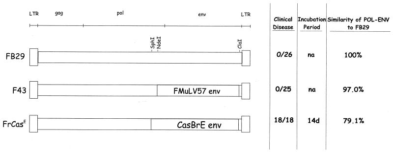
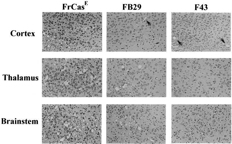
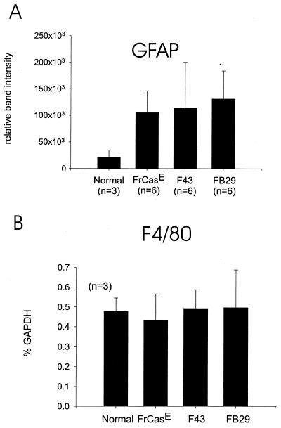
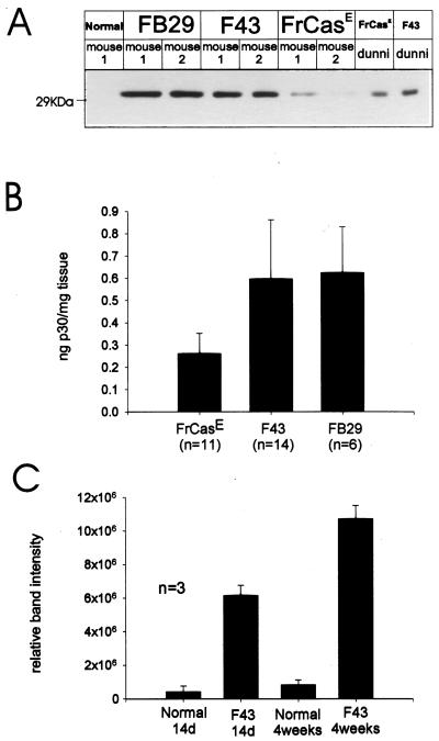
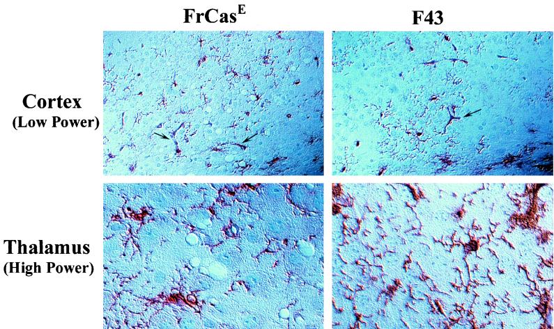
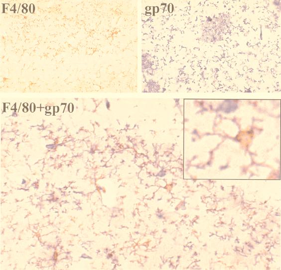
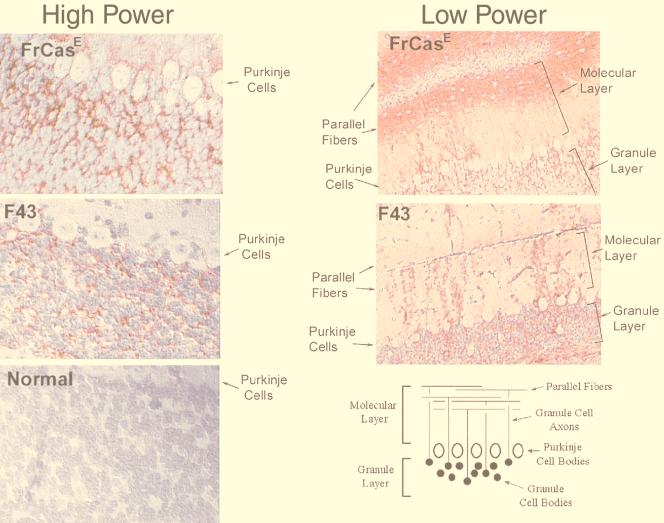
The chimeric murine oncornavirus FrCas(E) causes a rapidly progressive noninflammatory spongiform encephalomyelopathy after neonatal inoculation. The virus was constructed by the introduction of pol-env sequences from the wild mouse virus CasBrE into the genome of a neuroinvasive but nonneurovirulent strain of Friend murine leukemia virus (FMuLV), FB29. Although the brain infection by FrCas(E) as well as that by other neurovirulent murine retroviruses has been described in detail, little attention has been paid to the neuroinvasive but nonneurovirulent viruses. The purpose of the present study was to compare brain infection by FrCas(E) with that by FB29 and another nonneurovirulent virus, F43, which contains pol-env sequences from FMuLV 57. Both FB29 and F43 infected the same spectrum of cell types in the brain as that infected by FrCas(E), including endothelial cells, microglia, and populations of neurons which divide postnatally. Viral burdens achieved by the two nonneurovirulent viruses in the brain were actually higher than that of FrCas(E). The widespread infection of microglia by the two nonneurovirulent viruses is notable because it is infection of these cells by FrCas(E) which is thought to be a critical determinant of its neuropathogenicity. These results indicate that although the sequence of the envelope gene determines neurovirulence, this effect appears to operate through a mechanism which does not influence either viral tropism or viral burden in the brain. Although all three viruses exhibited similar tropism for granule neurons in the cerebellar cortex, there was a striking difference in the distribution of envelope proteins in those cells in vivo. The FrCas(E) envelope protein accumulated in terminal axons, whereas those of FB29 and F43 remained predominantly in the cell bodies. These observations suggest that differences in the intracellular sorting of these proteins may exist and that these differences appear to correlate with neurovirulence.


Figures

FIG. 1
FIG. 1
Schematic diagram of viral genomes showing the boundaries of the pol-env sequences introduced into the genome of FB29. The locations of viral genes are shown above. The long terminal repeats (LTRs), the gag genes, and the majority of the pol genes of all three viruses are identical, being derived from FB29. The pol-env sequences from FMuLV 57 and CasBrE introduced into the chimeric viruses F43 and FrCasE respectively, have different 5′ boundaries. However, there are no differences between FB29 and F43 in the pol coding sequence between SphI and NdeI. Shown to the right are results of a typical experiment, in which neonates were inoculated with the respective viruses and observed for evidence of clinical neurologic disease. Incubation period refers to the day after inoculation when the first signs of disease were noted. For FB29 and F43 this observation period could not be extended beyond 3 months because of the onset of erythroleukemia. na, not applicable.
FIG. 2
FIG. 2
Histopathology induced by FrCasE, FB29, and F43. Photomicrographs show hematoxylin- and eosin-stained sections through the deep cerebral cortex, thalamus, and the brain stem at the level of the vestibular nucleus. Mice were sacrificed at 16 days p.i., a time when the FrCasE-infected mice exhibited severe tremulous paralysis. Note that FrCasE induced spongiosis in all three brain regions. FB29 induced lesions of similar appearance in the thalamus and brain stem, but the spongiosis was minimal in extent. F43 caused only rare spongiform lesions in the brain stem, and this was an inconsistent finding. The large holes in the FB29 and F43 cortices (arrows) are blood vessels. Magnification before enlargement, ×135.
FIG. 3
FIG. 3
Quantification of markers of glial activation in mice 16 days after inoculation of FrCasE, F43, or FB29, a time point at which all FrCasE-inoculated mice exhibited severe neurologic disease. Upregulation of GFAP (A) is a marker of astrocytic activation (12) and was measured by semiquantitative Western blotting. Bands developed by chemiluminscence were quantified after digitization with ImageQuant (Molecular Dynamics) software and are expressed in terms of volume as a measure of relative signal strength. There was a significant difference (P = 0.01) between the infected mice (grouped together) and the uninoculated controls, but no significant differences between the infected groups were found. Upregulation of F4/80 (B) is a marker of microglial activation (2) and was measured at the mRNA level by an RNase protection assay. Data is expressed as percentage of the signal from the “housekeeping” gene encoding GAPDH. There was no evidence for upregulation of F4/80 in any of the infected mice.
FIG. 4
FIG. 4
Measurements of viral burden in the brain. Mice were infected intraperitoneally as neonates and sacrificed at 14 days p.i. when clinical signs of tremor and paralysis had appeared in the FrCasE-inoculated mice. Ten percent brain homogenates were analyzed for capsid protein (p30) either by polyacrylamide gel electrophoresis and immunoblot analysis with rabbit anti-p30 antiserum (A) or by antigen capture ELISA (B). Immunoblots were performed on two mice per group and are shown with positive controls on the right from extracts of M. dunni cells infected with either FrCasE or F43. ELISA was carried out on larger numbers of mice, and results were expressed as means ±1 standard deviation in units of nanograms of p30 per milligram of wet brain. Results indicated that both of the nonneurovirulent viruses (FB29 and F43) exhibited higher viral burdens in the brain than the neurovirulent virus (FrCasE). Immunoblot analysis (C) of brain extracts probed with anti-p30 antiserum compares the signal strengths for samples from mice inoculated with F43 at 2 weeks p.i. with that at 4 weeks p.i. Bands were quantified after digitization as described in the legend to Fig. 3.
FIG. 5
FIG. 5
Immunohistochemcial staining of infected cells in the brains of FrCasE- and F43-infected mice 16 days p.i. Sections of paraffin-embedded tissue were subjected to heat-induced antigen retrieval and stained with goat anti-gp70. The substrate was 3-amino-9-ethyl-carbazole, which yields a red color, and the sections were counterstained with hematoxylin. Sections were illuminated by differential-interference contrast. Morphologically, two types of cellular elements are seen to express viral gp70, cells associated with tubular and sometimes branched vascular structures (arrows) and highly arborized glial elements. The large nuclei which stain in the background are predominantly neurons. These photomicrographs illustrate the apparent lack of difference in the cell types infected by neurovirulent (FrCasE) and nonneurovirulent (F43) viruses. Magnifications before enlargement: low power, ×50; high power, ×100.
FIG. 6
FIG. 6
Colocalization of viral envelope protein and the microglial marker F4/80 in mice infected with F43. Sequential frozen sections were stained with either rabbit anti-F4/80, goat anti-gp70, or both (see Materials and Methods). The substrate for F4/80 was DAB, which produces an orange-brown color, and the substrate for anti-gp70 was Vector Elite, which produces a blue color. The sections viewed here are through the hippocampus and demonstrate extensive colocalization of the two substrates in the same highly arborized cells, identifying them as microglia. The patches of staining seen in the gp70 panel are characteristically seen in the superficial layers of the cerebral cortex and the hippocampus (shown here) for all three of the viruses examined in this study. These patches appear not to be cell associated though their nature is currently not known. Magnification before enlargement, ×100.
FIG. 7
FIG. 7
Difference in the localization of FrCasE and F43 envelope proteins in the cerebellum. Photomicrographs show paraffin sections through the cerebellar cortex 14 days p.i., stained with goat anti-gp70 and AEC (red color) as the substrate. (Left) Granule layers of the cerebellar cortices of mice inoculated with FrCasE, F43, and an uninoculated control. Like FrCasE (23), F43 also infected neurons in that layer, as revealed by the red stain. There is a slight difference in the hematoxylin counterstain between the panels (original magnification, ×100). (Right) Low-power views (original magnification, ×25) of the full thickness of the cerebellar cortex, revealing the distribution of the respective envelope proteins in the molecular layer. The schematic at the bottom shows the cellular anatomy of the cerebellar cortex. The axons of granule neurons course through the row of Purkinje cells into the molecular layer, where they bifurcate and extend laterally forming the parallel fibers. Whereas there is extensive staining of the FrCasE envelope protein in the parallel-fiber layer, this was not observed for F43. Instead one observes in F43-inoculated mice focal staining in the molecular layer in a radial pattern, suggesting that the envelope protein reached the proximal but not distal axons.

Similar articles

Cited by

References

    1. Albritton L M, Tseng L, Scadden D, Cunningham J M. A putative murine ecotropic retrovirus receptor gene encodes a multiple membrane-spanning protein and confers susceptibility to virus infection. Cell. 1989;57:659–666. - PubMed
    1. Andersson P B, Perry V H, Gordon S. The kinetics and morphological characteristics of the macrophage-microglial response to kainic acid-induced neuronal degeneration. Neuroscience. 1991;42:201–214. - PubMed
    1. Battini J-L, Heard J M, Danos O. Receptor choice determinants in the envelope glycoproteins of amphotropic, xenotropic, and polytropic murine leukemia viruses. J Virol. 1992;66:1468–1475. - PMC - PubMed
    1. Chesebro B, Britt W, Evans L, Wehrly K, Nishio J, Cloyd M. Characterization of monoclonal antibodies reactive with murine leukemia viruses: use in analysis of strains of Friend MCF and Friend ecotropic murine leukemia virus. Virology. 1983;127:134–148. - PubMed
    1. Czub M, Czub S, McAtee F J, Portis J L. Age-dependent resistance to murine retrovirus-induced spongiform neurodegeneration results from central nervous system-specific restriction of virus replication. J Virol. 1991;65:2539–2544. - PMC - PubMed

LinkOut - more resources