

doi: 10.1128/jvi.74.1.580-583.2000.
Identification of a bovine coronavirus packaging signal
Affiliations
- PMID: 10590153
- PMCID: PMC111575
- DOI: 10.1128/jvi.74.1.580-583.2000
Item in Clipboard
Identification of a bovine coronavirus packaging signal
J Virol.
2000 Jan.
Abstract
A region of the bovine coronavirus (BCV) genome that functions as a packaging signal has been cloned. The 291-nucleotide clone shares 72% homology with the region of mouse hepatitis coronavirus (MHV) gene 1b that contains the packaging signal. RNA transcripts were packaged into both BCV and MHV virions when the cloned region was appended to a noncoronavirus RNA. This is the first identification of a BCV packaging signal. The data demonstrate that the BCV genome contains a sequence that is conserved at both the sequence and functional levels, thus broadening our insight into coronavirus packaging.
Figures
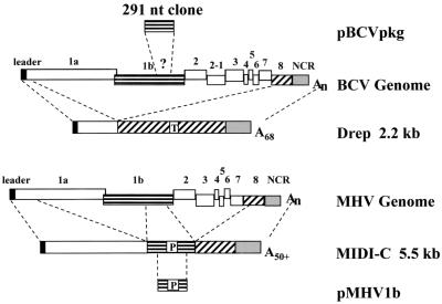
Comparison of the BCV and MHV strain A59 genomes and representative defective genomes. Drep is a cloned BCV defective RNA (6), and MIDI-C is a cloned MHV strain A59 DI RNA (7). The various parts of the genome that are included in each defective genome are indicated by dashed lines extending from the schematic of the parental genome. Drep includes a 30-nt reporter sequence (T) derived from the TGEV N gene. MIDI-C includes a known packaging signal (P). The approximate location of the packaging signal in the MHV genome (P) and the assumed position of the 291-nt cloned region of BCV (?) are indicated. Schematic representations of the 291-nt BCV RT-PCR and 312-nt MHV PCR products included in plasmids pBCVpkg and pMHV1b, respectively, are shown. NCR, noncoding region.
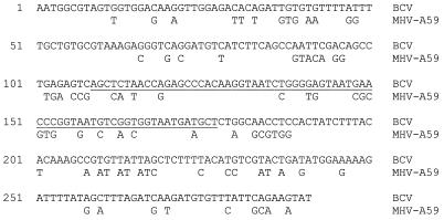
Sequence of the cloned BCV region and alignment with the homologous region from the MHV strain A59 genome. The region of the BCV clone that is homologous to the MHV packaging signal is underlined. Only nonidentical nucleotides are shown for MHV.
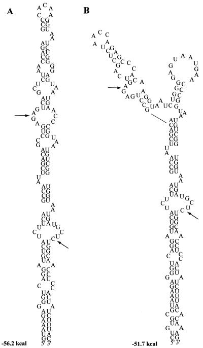
Predicted secondary RNA structures of the cloned BCV region. Predictions were made in the context of both the 190-nt (nt 42 to 232 in Fig. 2) and 291-nt (nt 1 to 291) RNAs. Only nt 59 to 200 (Fig. 2) are shown for simplicity. Arrows indicate the position of the 69-nt region that shares homology with the MHV packaging signal.
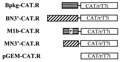
Schematic representation of constructs used to test the ability of the BCV clone to function as a packaging signal. All coronavirus sequences were placed under the control of the T7 promoter. A cassette containing the CAT gene, the hepatitis delta virus ribozyme (r), and the T7 terminator (T7t) was cloned at the 3′ end of each coronavirus sequence.
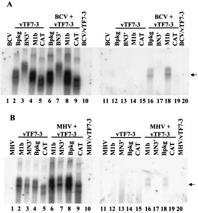
Northern blot analysis of expressed RNA transcripts packaged by BCV (A) and MHV strain A59 (B). Plasmid DNAs were transfected into vTF7-3-infected cells (lanes 2 to 9 and 12 to 19). Both intracellular RNAs (lanes 1 to 10) and extracellular virion RNAs (lanes 11 to 20) were analyzed by Northern blotting after cells were mock infected (lanes 2 to 5 and 12 to 15) or infected (lanes 6 to 9 and 16 to 19) with BCV (A) or MHV strain A59 (B). Extracellular media were treated with both DNase and RNase prior to isolation of virions. RNAs were analyzed by using a CAT-specific riboprobe. Arrows indicate the position of the packaged Bpkg-CAT and M1b-CAT RNAs.
References
-
- Bredenbeek B J, Pachuk C J, Notern A F H, Charite J, Luytjes W, Weiss S R, Spaan W J M. The primary structure and expression of the 2 open reading frame of the polymerase gene of the coronavirus MHV-A59—a highly conserved polymerase is expressed by an efficient ribosomal frame-shifting mechanism. Nucleic Acids Res. 1990;18:1825–1832. - PMC - PubMed
Publication types
MeSH terms
Substances
Associated data
- Actions
Grants and funding
LinkOut - more resources
Full Text Sources
Other Literature Sources
