. 2000 Jan;20(1):61-9.
doi: 10.1128/MCB.20.1.61-69.2000.

Histone H1 is dispensable for methylation-associated gene silencing in Ascobolus immersus and essential for long life span

Affiliations

Histone H1 is dispensable for methylation-associated gene silencing in Ascobolus immersus and essential for long life span

J L Barra et al. Mol Cell Biol. 2000 Jan.

Abstract

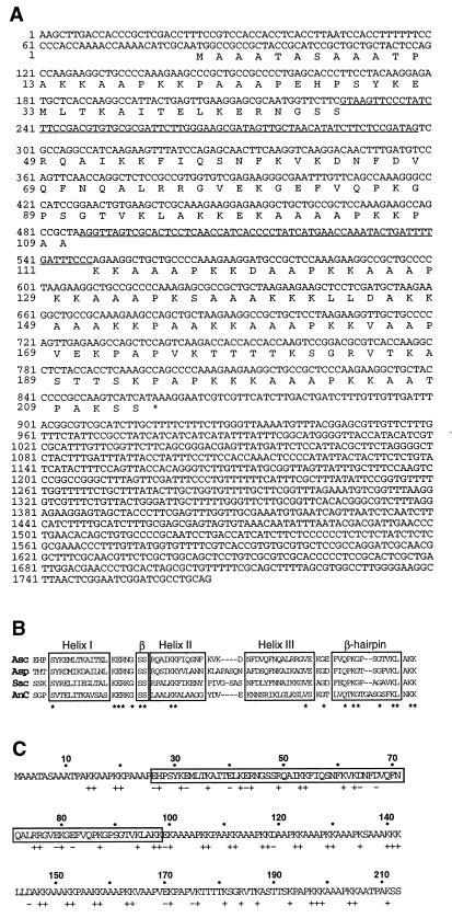
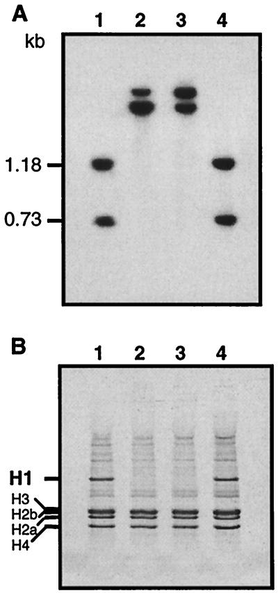
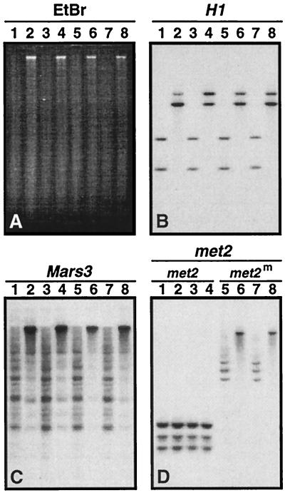
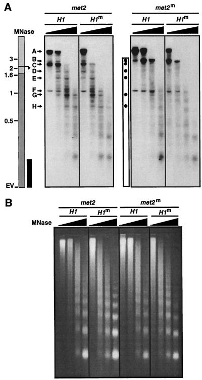
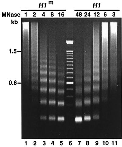
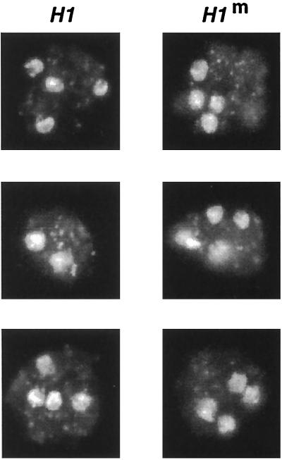
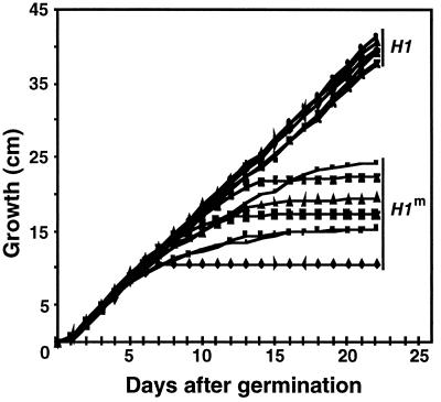
A gene encoding a protein that shows sequence similarity with the histone H1 family only was cloned in Ascobolus immersus. The deduced peptide sequence presents the characteristic three-domain structure of metazoan linker histones, with a central globular region, an N-terminal tail, and a long positively charged C-terminal tail. By constructing an artificial duplication of this gene, named H1, it was possible to methylate and silence it by the MIP (methylation induced premeiotically) process. This resulted in the complete loss of the Ascobolus H1 histone. Mutant strains lacking H1 displayed normal methylation-associated gene silencing, underwent MIP, and showed the same methylation-associated chromatin modifications as did wild-type strains. However, they displayed an increased accessibility of micrococcal nuclease to chromatin, whether DNA was methylated or not, and exhibited a hypermethylation of the methylated genome compartment. These features are taken to imply that Ascobolus H1 histone is a ubiquitous component of chromatin which plays no role in methylation-associated gene silencing. Mutant strains lacking histone H1 reproduced normally through sexual crosses and displayed normal early vegetative growth. However, between 6 and 13 days after germination, they abruptly and consistently stopped growing, indicating that Ascobolus H1 histone is necessary for long life span. This constitutes the first observation of a physiologically important phenotype associated with the loss of H1.


Figures

FIG. 1
FIG. 1
Primary structure of histone H1 from Ascobolus. (A) Nucleotide and derived amino acid sequences of the H1 gene. The entire 1.7-kb HindIII-PstI fragment is shown. The two introns are underlined. (B) Comparison of the amino acid sequence of the globular domain of Ascobolus H1 (Asc) with the globular domains of H1 from A. nidulans (Asp) and S. cerevisae (Sac; the first globular domain is shown) and of the H1 consensus sequence resulting from the comparison of 30 animal H1 sequences (58). Boxes indicate the regions identified as α helix and β sheet in the H5 crystal structure (39). Asterisks indicate the conserved amino acids. (C) Tripartite organization of the Ascobolus H1 protein. The box indicates the globular domain flanked by the N- and C-terminal tails. Positions of basic (+) and acidic (−) residues are indicated.
FIG. 2
FIG. 2
Methylation and silencing of the H1 gene. (A) Southern analysis of methylation in four strains derived from the H1-duplicated strain and harboring only the resident H1 copy. Sau3AI DNA digests were probed with the cloned 1.7-kb HindIII-PstI fragment carrying the H1 gene (this work). Two strains (lanes 1 and 4) harbored the unmethylated allele, as revealed by the presence of the 1,184- and 728-bp expected fragments. The two other strains (lanes 2 and 3) harbored the methylated allele, as revealed by the presence of larger fragments. (B) SDS-PAGE of H2SO4-soluble proteins extracted from nuclei of the same strains as in panel A. The bands corresponding to H1 and to the core histones are indicated.
FIG. 3
FIG. 3
Hypermethylation in strains lacking histone H1. (A) Ethidium bromide staining of the gel used for Southern analyses of the Sau3AI DNA digests shown in panels B to D. As determined from panel B, strains analyzed in lanes 1, 3, 5, and 7 harbored the native unmethylated H1 allele, and strains analyzed in lanes 2, 4, 6, and 8 harbored the methylated allele. The gel was probed with H1 (B), Mars3 (C), and met2 (D). The H1 probe was as in Fig. 2. The Mars3 probe corresponded to the EcoRI fragment carrying Mars3 in plasmid pCG57 (16). The met2 probe was a PCR product corresponding to the 2.9-kb HincII-BglII fragment carrying the met2 gene (14). Strains 5 to 8 had inherited a silenced allele of met2, while strains 1 to 4 expressed this gene. The largest met2 Sau3AI fragments from strains 5 and 7 are hardly visible on the autoradiograph presented in panel D.
FIG. 4
FIG. 4
Comparison of the methylation-associated chromatin modifications in H1-silenced and wild-type strains. (A) MNase analysis of the chromatin of the met2 gene was performed in two strains harboring the unmethylated allele of met2 (met2) and two strains harboring the methylated allele of this gene (met2m). One strain of each type harbored the silenced allele of H1 (H1m); the other harbored the wild-type allele (H1). Protoplasts were incubated with increasing amounts of MNase and subjected to indirect end-labeling analysis. Samples were loaded on the same gel. The long vertical box at the left represents the met2 gene, with the transcription start site (arrow), ORF (dark gray box), position of the EcoRV site (EV), and size markers (in kilobases) indicated. The black vertical box indicates the probe used for hybridization. A to H indicate the eight major bands obtained when met2 was unmethylated (met2,H1; met2,H1m). Band A corresponds to the EcoRV fragment carrying the met2 gene. The white vertical box indicates the methylated region of met2. Black dots indicate the positions of bands that changed when met2 was methylated (metm,H1; met2m,H1m). (B) Ethidium bromide staining of the gel used for hybridization shown in panel A.
FIG. 5
FIG. 5
Nucleosome repeat ladder obtained after chromatin digestion with MNase of one strain harboring the silenced allele of H1 (H1m) and one strain harboring the wild-type allele (H1). In the central lane is the size marker ladder (100-bp ladder; GIBCO BRL). The relative amounts of MNase used are indicated above the lanes.
FIG. 6
FIG. 6
Comparison of DAPI-stained nuclei in protoplasts from the wild-type strain (H1) and strains lacking H1 (H1m).
FIG. 7
FIG. 7
Comparison of growth rates of six strains harboring the silenced copy of the H1 gene (H1m) and six wild-type strains (H1).

References

    1. Allan J, Mitchell T, Harborne N, Bohm L, Crane-Robinson C. Roles of H1 domains in determining higher order chromatin structure and H1 location. J Mol Biol. 1986;187:591–601. - PubMed
    1. Antequera F, Boyes J, Bird A P. High levels of de novo methylation and altered chromatin structure at CpG islands in cell lines. Cell. 1990;62:503–514. - PubMed
    1. Ball D J, Gross D S, Garrard W T. 5-Methylcytosine is localized in nucleosomes that contain histone H1. Proc Natl Acad Sci USA. 1983;80:5490–5494. - PMC - PubMed
    1. Bouvet P, Dimitrov S, Wolffe A P. Specific regulation of Xenopus chromosomal 5S rRNA gene transcription in vivo by histone H1. Genes Dev. 1994;8:1147–1159. - PubMed
    1. Campoy F J, Meehan R R, McKay S, Nixon J, Bird A P. Binding of histone H1 to DNA is indifferent to methylation at CpG sequences. J Biol Chem. 1995;270:26473–26481. - PubMed

Publication types