. 2000 Jan;20(1):91-103.
doi: 10.1128/MCB.20.1.91-103.2000.

Auto-inhibition and partner proteins, core-binding factor beta (CBFbeta) and Ets-1, modulate DNA binding by CBFalpha2 (AML1)

Affiliations

Auto-inhibition and partner proteins, core-binding factor beta (CBFbeta) and Ets-1, modulate DNA binding by CBFalpha2 (AML1)

T L Gu et al. Mol Cell Biol. 2000 Jan.

Abstract

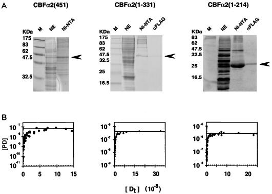
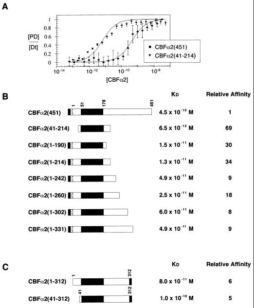
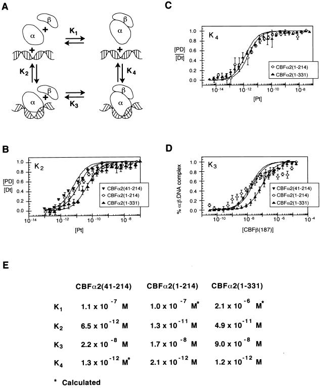
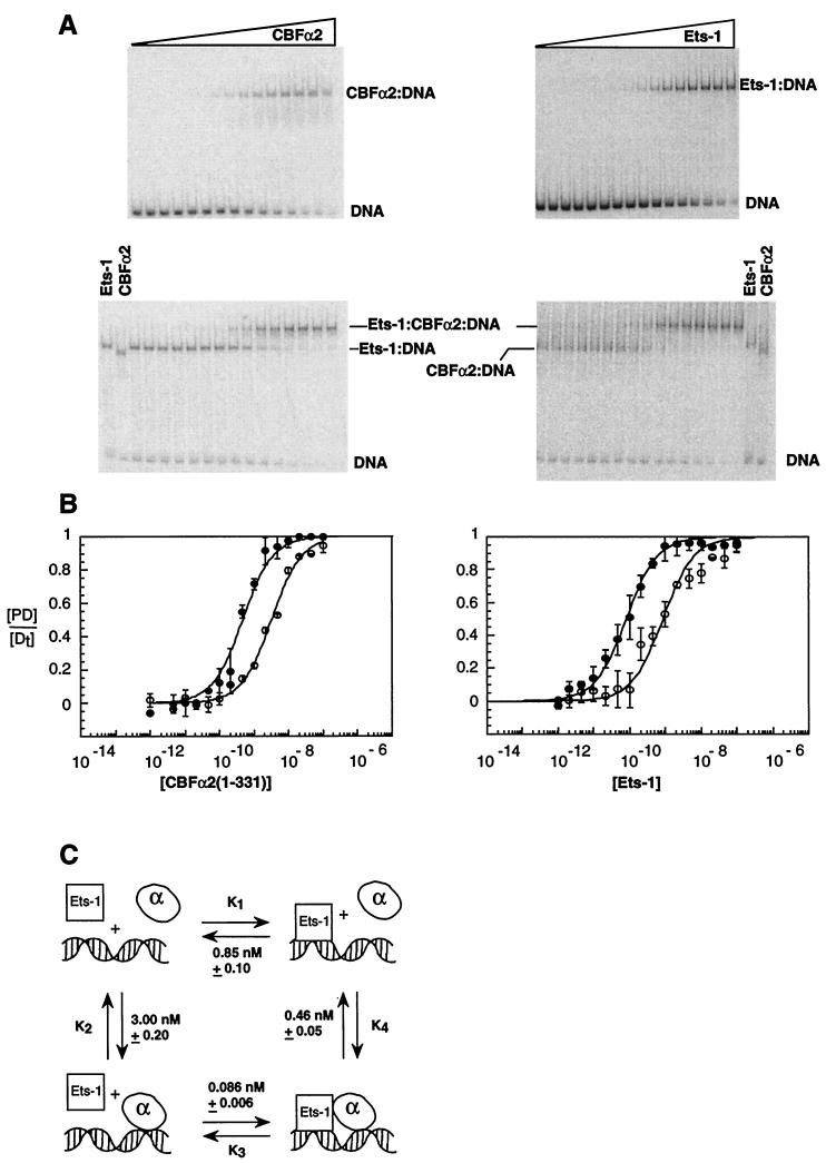
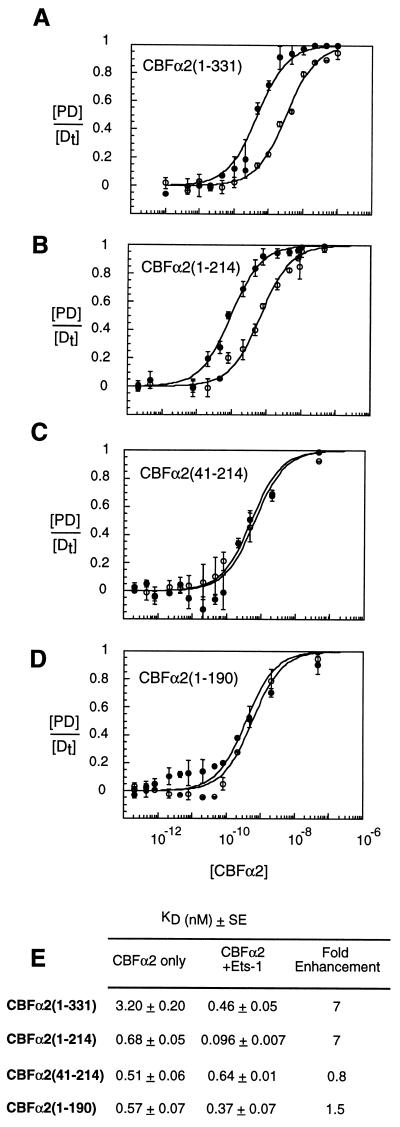
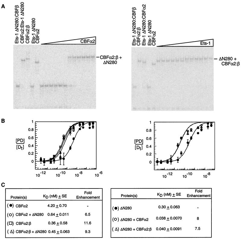
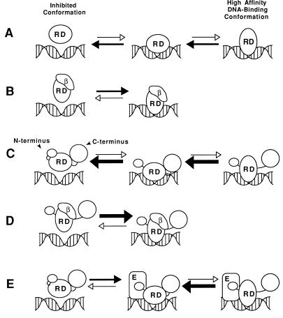
Core-binding factor alpha2 (CBFalpha2; otherwise known as AML1 or PEBP2alphaB) is a DNA-binding subunit in the family of core-binding factors (CBFs), heterodimeric transcription factors that play pivotal roles in multiple developmental processes in mammals, including hematopoiesis and bone development. The Runt domain in CBFalpha2 (amino acids 51 to 178) mediates DNA binding and heterodimerization with the non-DNA-binding CBFbeta subunit. Both the CBFbeta subunit and the DNA-binding protein Ets-1 stimulate DNA binding by the CBFalpha2 protein. Here we quantify and compare the extent of cooperativity between CBFalpha2, CBFbeta, and Ets-1. We also identify auto-inhibitory sequences within CBFalpha2 and sequences that modulate its interactions with CBFbeta and Ets-1. We show that sequences in the CBFalpha2 Runt domain and sequences C terminal to amino acid 214 inhibit DNA binding. Sequences C terminal to amino acid 214 also inhibit heterodimerization with the non-DNA-binding CBFbeta subunit, particularly heterodimerization off DNA. CBFbeta rescinds the intramolecular inhibition of CBFalpha2, stimulating DNA binding approximately 40-fold. In comparison, Ets-1 stimulates CBFalpha2 DNA binding 7- to 10-fold. Although the Runt domain alone is sufficient for heterodimerization with CBFbeta, sequences N terminal to amino acid 41 and between amino acids 190 and 214 are required for cooperative DNA binding with Ets-1. Cooperative DNA binding with Ets-1 is less pronounced with the CBFalpha2-CBFbeta heterodimer than with CBFalpha2 alone. These analyses demonstrate that CBFalpha2 is subject to both negative regulation by intramolecular interactions, and positive regulation by two alternative partnerships.


Figures

FIG. 1
FIG. 1
Expression and purification of CBFα2. (A) Coomassie blue-stained sodium dodecyl sulfate-polyacrylamide gel displaying fractions from each step of the purification for CBFα2(451) and two truncated derivatives, CBFα2(1-331) and CBFα2(1-214). Lanes: M, molecular weight markers; NE, unfractionated nuclear extract; Ni-NTA, eluate from the Ni-NTA column; αFLAG, eluate from the anti-FLAG monoclonal antibody column. Arrows indicate expected position of the CBFα2 bands. (B) Activities of CBFα2 proteins quantified by DNA titration in an EMSA. Concentrations (molar) of protein-DNA complex [PD] versus total input DNA [Dt] are plotted.
FIG. 2
FIG. 2
Modulation of CBFα2 DNA binding by C-terminal sequences. (A) Equilibrium DNA binding studies of full-length CBFα2(451) and CBFα2(41-214) were performed by EMSA and used to generate DNA binding curves. Data from at least three experiments provide mean and standard error for each data point. KD values were obtained by curve fitting as described in Materials and Methods. (B) Summary of equilibrium dissociation constants for truncated CBFα2. The black rectangle in the schematic diagram of CBFα2 represents the DNA-binding Runt domain. The gray and stippled boxes represent the H6 and FLAG tags, respectively. Relative affinity was calculated as the ratio of mutant affinity to the affinity of CBFα2(451). (C) Summary of equilibrium dissociation constants for CBFα2 proteins tagged at amino acid 312 with H6 (gray box).
FIG. 3
FIG. 3
Thermodynamic box describing interactions between CBFα2, CBFβ, and DNA. (A) Schematic diagram of the potential interactions between CBFα2 (α), CBFβ (β), and DNA. The modeled bend in DNA induced by the Runt domain is suggested by both circular permutation analysis and circular dichroism spectroscopy (; Crute et al., submitted). (B) Equilibrium dissociation constants (K2) of CBFα2(41-214), CBFα2(1-214), and CBFα2(1-331) for DNA. Data from at least three experiments are presented. Standard errors are 1.1 × 10⁻¹² M, 2.1 × 10⁻¹² M, and 7.1 × 10⁻¹² M, respectively. (C) Equilibrium dissociation constants (K4) of CBFα2-CBFβ heterodimers for DNA. Standard errors are 3.9 × 10⁻¹³ M for CBFα2(1-214) and 1.8 × 10⁻¹³ M for CBFα2(1-331). (D) Equilibrium dissociation constants (K3) of CBFβ for CBFα2-DNA complexes. Data represent at least three experiments. Standard errors are 3.2 × 10⁻⁹ M, 1.5 × 10⁻⁹ M, and 3.5 × 10⁻⁹ M for CBFα2(41-214), CBFα2(1-214), and CBFα2(1-331), respectively. (E) Summary of equilibrium dissociation constants K1, K2, K3, and K4. K4 for CBFα2(41-214) was not determined directly but calculated from K2K3 = K1K4. K1 for CBFα2(41-214) was determined independently (Crute et al., submitted).
FIG. 4
FIG. 4
Ets-1 and CBFα2 bind DNA cooperatively. (A) EMSA of equilibrium DNA binding studies of CBFα2(1-331) titrated onto DNA alone or in the presence of Ets-1 (left) or Ets-1 titrated onto DNA alone and in the presence of CBFα2(1-331) (right). (B) Equilibrium DNA binding curves for CBFα2(1-331) (left) and Ets-1 (right); data from panel A. Symbols: ○, binary protein = DNA complexes; ●, ternary complexes. Equilibrium DNA binding curves display [PD/[Dt] as the mean (±standard error) of at least two independent experiments. (C) Thermodynamic box depicting potential interactions between Ets-1, CBFα2, and DNA. Equilibrium dissociation constants were obtained from panels A and B. KD values and standard error were obtained from the curve fit of means as described in Materials and Methods.
FIG. 5
FIG. 5
Sequences in CBFα2 required for cooperative DNA binding with Ets-1. Equilibrium DNA binding studies were performed by EMSA with truncated CBFα2 proteins in the absence (open circles) or presence (closed circles) of Ets-1 (A to D). Equilibrium DNA binding curves display [PD]/[Dt] as the mean (±standard error) of at least two independent experiments. (E) Summary of equilibrium dissociation constants derived from binding curves in panels A to D. KD values and standard error were obtained from the curve fit of means as described in Materials and Methods.
FIG. 6
FIG. 6
DNA-binding enhancement by Ets-1 and CBFβ is neither additive nor synergistic. (A) EMSA of equilibrium DNA binding studies of CBFα2(1-331) titrated onto DNA saturated with Ets-1ΔN280 in the presence of CBFβ protein (left) and of Ets-1ΔN280 titrated onto DNA saturated with the CBFα2-CBFβ heterodimer (right). Control lanes to the left of each panel document the position of each of the protein-DNA complexes. (B) Equilibrium DNA binding curves for CBFα2(1-331) (left) and Ets-1ΔN280 (right). The identity of each curve is indicated in panel C. (C) Summary of equilibrium dissociation constants. Relative binding affinities (fold enhancement) compare KD values for multiprotein-DNA complexes to those obtained from DNA binding studies of Ets-1 and CBFα2 in isolation. KD values are presented as the mean (±standard error) of at least two independent experiments.
FIG. 7
FIG. 7
Models for interactions between CBFα2, CBFβ, and Ets-1. (A) The Runt domain (RD) is in equilibrium between a high- and low-affinity DNA-binding conformation. (B) Heterodimerization with CBFβ (β) locks the Runt domain into its high-affinity DNA-binding conformation, shifting the DNA-binding equilibrium to the right. (C) C-terminal inhibitory sequences in CBFα2 further shift the equilibrium of the Runt domain toward its low-affinity DNA-binding conformation and mask the CBFβ heterodimerization surface. Association of the C-terminal inhibitory sequences to the Runt domain is destabilized when CBFα2 is bound to DNA. Dissociation of the inhibitory sequences unmasks the CBFβ binding surface on the Runt domain. (D) The high-affinity DNA-binding conformation of the Runt domain is stabilized by the CBFβ subunit. Association of the C-terminal inhibitory sequences to the Runt domain is also directly inhibited by the CBFβ subunit, which masks the interaction site. The DNA-binding affinity of this complex is the same as that of the Runt domain-CBFβ complex in panel B. (E) Binding of CBFα2 to DNA exposes the Ets-1 interaction surface, which includes (but is not restricted to) sequences N terminal to the Runt domain. Tethering of Ets-1 to CBFα2 on the DNA increases the likelihood of a productive binding event, resulting in increased affinity. Ets-1 does not mask the Runt domain surface to which CBFβ and the C-terminal inhibitory domain bind. Conformational changes in the Ets-1 protein itself are not depicted in this diagram (see the accompanying report [20]).
FIG. 8
FIG. 8
Summary of CBFα2(451) functional domains. Shown are boundaries of the DNA-binding and heterodimerization domains as defined by Kagoshima et al. (30). Autoinhibition of both DNA binding and heterodimerization maps to the C-terminal half of the protein. RD, Runt domain.

References

    1. Adja N, Stacy T, Speck N A, Liu P P. The leukemic protein CBFβ-SMMHC sequesters CBFα2 into cytoskeletal filaments and aggregates. Mol Cell Biol. 1998;18:7432–7443. - PMC - PubMed
    1. Albagli O, Klaes A, Ferreira E, Leprince D, Klambt C. Function of ets genes is conserved between vertebrates and Drosophila. Mech Dev. 1996;59:29–40. - PubMed
    1. Bae S-C, Ogawa E, Maruyama M, Oka H, Satake M, Shigesada K, Jenkins N A, Gilbert D J, Copeland N G, Ito Y. PEBP2αB/mouse AML1 consists of multiple isoforms that possess differential transactivation potentials. Mol Cell Biol. 1994;14:3242–3252. - PMC - PubMed
    1. Bae S-C, Takahashi E, Zhang Y W, Ogawa E, Shigesada K, Namba Y, Satake M, Ito Y. Cloning, mapping and expression of PEBP2αC, a third gene encoding the mammalian Runt domain. Gene. 1995;159:245–248. - PubMed
    1. Bae S C, Yamaguchi-Iwai Y, Ogawa E, Maruyama M, Inuzuka M, Kagoshima H, Shigesada K, Satake M, Ito Y. Isolation of PEBP2αB cDNA representing the mouse homolog of human acute myeloid leukemia gene, AML1. Oncogene. 1993;8:809–814. - PubMed

Publication types

LinkOut - more resources