. 2000 Jan;20(1):113-25.
doi: 10.1128/MCB.20.1.113-125.2000.

Alleviation of human papillomavirus E2-mediated transcriptional repression via formation of a TATA binding protein (or TFIID)-TFIIB-RNA polymerase II-TFIIF preinitiation complex

Affiliations

Alleviation of human papillomavirus E2-mediated transcriptional repression via formation of a TATA binding protein (or TFIID)-TFIIB-RNA polymerase II-TFIIF preinitiation complex

S Y Hou et al. Mol Cell Biol. 2000 Jan.

Abstract

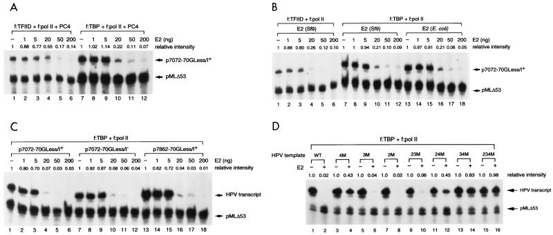
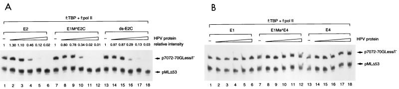
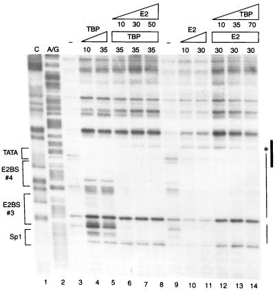
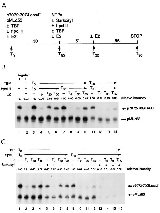
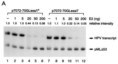
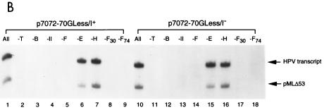
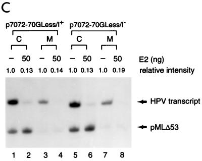
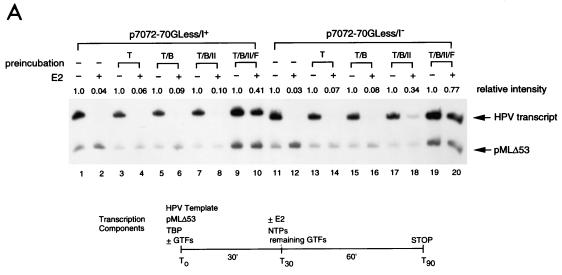
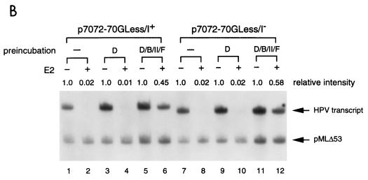
Transcription in human papillomaviruses (HPVs) is mainly regulated by cellular transcription factors and virus-encoded E2 proteins that act as sequence-specific DNA-binding proteins. Although the functions of E2 as a transcriptional activator and a repressor have been well documented, the role of cellular factors involved in E2-mediated regulation of the HPV promoters and the mechanism by which E2 modulates viral gene expression remain unclear. Using reconstituted cell-free transcription systems, we found that cellular enhancer-binding factors and general cofactors, such as TAF(II)s, TFIIA, Mediator, and PC4, are not required for E2-mediated repression. Unlike other transcriptional repressors that function through recruitment of histone deacetylase or corepressor complexes, HPV E2 is able to directly target components of the general transcription machinery to exert its repressor activity on the natural HPV E6 promoter. Interestingly, preincubation of TATA binding protein (TBP) or TFIID with HPV template is not sufficient to overcome E2-mediated repression, which can be alleviated only via formation of a minimal TBP (or TFIID)-TFIIB-RNA polymerase II-TFIIF preinitiation complex. Our data therefore indicate that E2 does not simply work by displacing TBP or TFIID from binding to the adjacent TATA box. Instead, E2 appears to function as an active repressor that directly inhibits HPV transcription at steps after TATA recognition by TBP or TFIID.


Figures

FIG. 1
FIG. 1
Recapitulation of E2-mediated transcriptional regulation of the homologous E6 promoter in vitro. (A) HPV-11 E2, E1, and E4 family proteins. Numbers next to carets indicate the nucleotide positions of exon boundaries adjacent to splice donors and acceptors, whereas numbers at the beginning and end of each box are the first and last nucleotides of individual open reading frames. The E2, E1, and E4 open reading frames are shown as white, gray, and black boxes, respectively. The apparent molecular mass (MW) of each protein, estimated by sodium dodecyl sulfate-polyacrylamide gel electrophoresis, is also listed on the right. (B) Purified recombinant HPV-11 proteins. Coomassie blue staining of FLAG-tagged HPV-11 proteins purified from either bacteria (lanes 1 to 6) or Sf9 insect cells (lanes 8 to 12) by immunoaffinity purification and peptide elution methods (see Materials and Methods) is shown. The positions of HPV-11 proteins separated by sodium dodecyl sulfate–15% polyacrylamide gel electrophoresis are indicated on the left. Prestained protein size markers (in kilodaltons; from GIBCO-BRL) are depicted on the right. (C) Transcriptional regulation of the HPV-11 E6 promoter by homologous E2 protein. In vitro transcription was performed in HeLa nuclear extracts (N.E.) with pGL7072-161, which contains the HPV-11 URR spanning nucleotides 7072 to 7933/1 to 161, and an HIV-1 internal control template, pHIV+58, in the absence (−) or presence of increasing amounts of baculovirus-expressed HPV-11 E2 protein. Transcripts derived from the HPV-11 and HIV-1 templates are indicated, respectively, by arrows. Relative intensity is defined as the signal intensity quantitated by PhosphorImager (Molecular Dynamics) from the HPV template relative to that performed in the absence of E2. (D) Mapping of the HPV-11 E6 promoter start site. In vitro transcription was performed with HeLa nuclear extracts with pGL7072-161, and the RNA synthesized was subjected to primer extension analysis as described in Materials and Methods. Different amounts of α-amanitin (in micrograms per milliliter) were also included in the reactions to score for pol II-specific transcripts. The DNA sequencing marker, prepared from the same primer and DNA template as employed for in vitro transcription, was used for the assignment of the transcription initiation site. The DNA sequence surrounding the transcription start site (+1) at P93, indicated by the arrow, and the TATA box of the E6 promoter are shown on the left.
FIG. 1
FIG. 1
Recapitulation of E2-mediated transcriptional regulation of the homologous E6 promoter in vitro. (A) HPV-11 E2, E1, and E4 family proteins. Numbers next to carets indicate the nucleotide positions of exon boundaries adjacent to splice donors and acceptors, whereas numbers at the beginning and end of each box are the first and last nucleotides of individual open reading frames. The E2, E1, and E4 open reading frames are shown as white, gray, and black boxes, respectively. The apparent molecular mass (MW) of each protein, estimated by sodium dodecyl sulfate-polyacrylamide gel electrophoresis, is also listed on the right. (B) Purified recombinant HPV-11 proteins. Coomassie blue staining of FLAG-tagged HPV-11 proteins purified from either bacteria (lanes 1 to 6) or Sf9 insect cells (lanes 8 to 12) by immunoaffinity purification and peptide elution methods (see Materials and Methods) is shown. The positions of HPV-11 proteins separated by sodium dodecyl sulfate–15% polyacrylamide gel electrophoresis are indicated on the left. Prestained protein size markers (in kilodaltons; from GIBCO-BRL) are depicted on the right. (C) Transcriptional regulation of the HPV-11 E6 promoter by homologous E2 protein. In vitro transcription was performed in HeLa nuclear extracts (N.E.) with pGL7072-161, which contains the HPV-11 URR spanning nucleotides 7072 to 7933/1 to 161, and an HIV-1 internal control template, pHIV+58, in the absence (−) or presence of increasing amounts of baculovirus-expressed HPV-11 E2 protein. Transcripts derived from the HPV-11 and HIV-1 templates are indicated, respectively, by arrows. Relative intensity is defined as the signal intensity quantitated by PhosphorImager (Molecular Dynamics) from the HPV template relative to that performed in the absence of E2. (D) Mapping of the HPV-11 E6 promoter start site. In vitro transcription was performed with HeLa nuclear extracts with pGL7072-161, and the RNA synthesized was subjected to primer extension analysis as described in Materials and Methods. Different amounts of α-amanitin (in micrograms per milliliter) were also included in the reactions to score for pol II-specific transcripts. The DNA sequencing marker, prepared from the same primer and DNA template as employed for in vitro transcription, was used for the assignment of the transcription initiation site. The DNA sequence surrounding the transcription start site (+1) at P93, indicated by the arrow, and the TATA box of the E6 promoter are shown on the left.
FIG. 2
FIG. 2
Use of G-less cassette templates for transcriptional analysis of HPV-11 E6 promoter activity. (A) HPV-11 G-less cassettes. Plasmids p7072-70GLess/I+ and p7072-70GLess/I contain the HPV-11 URR spanning nucleotides 7072 to 7933/1 to 70 linked to either a G-less cassette of 388 nucleotides preceded by the adenovirus MLP Inr element (I+) or another G-less cassette of 377 nucleotides without the MLP Inr (I). Plasmids p7862-70GLess/I+ and p7862-70GLess/I containing a truncated HPV-11 URR from nucleotide 7862 to nucleotide 70 without CEI and CEII were similarly linked to G-less cassettes with or without the MLP Inr. The compilation of cis-acting elements and trans-acting factors in the HPV-11 URR is mainly based on published information (28, 44, 72). The boundaries of CEI (28) and CEII (13) and the origin of replication (7) are indicated by brackets. (B) Start site mapping of HPV-11 and MLP G-less cassette templates. In vitro transcription reactions were performed with HeLa nuclear extracts with either an adenovirus MLP-driven G-less cassette template (pMLΔ53 [42]), HPV-11 p7072-70GLess/I+ and p7862-70GLess/I+ templates (both give the same start sites and are thus indicated as HPV/I+), or p7072-70GLess/I and p7862-70GLess/I templates (both give the same start sites and are thus indicated as HPV/I). The transcription start sites (arrows), MLP Inr (underlined), and TATA boxes (brackets) are shown on the left of each panel with intensities of relative start sites denoted by dots. (C) Comparison of nucleotide sequences surrounding the core promoter elements of the MLP and HPV templates. HPV-11 nucleotides are in boldface while vector or MLP sequences are in regular type. Brackets indicate the boundaries of the TATA boxes with lines above the sequence demarking the MLP initiator element. The promoter-proximal E2-binding sites (E2-BS 3 and E2-BS 4) are underlined, and the nucleotides (open arrows) corresponding to HPV-11 number designations are indicated in parentheses. (D) E2-mediated repression of the homologous E6 promoter. In vitro transcription was performed with HeLa nuclear extracts with 50 ng of each HPV and pMLΔ53 templates, in the absence (−) or presence of increasing amounts of baculovirus-expressed E2 protein.
FIG. 3
FIG. 3
E2-mediated repression of the HPV-11 E6 promoter in a reconstituted two-component transcription system. (A) TAFIIs are not required for E2-mediated repression. In vitro transcription was performed with TFIID-deficient pol II holoenzyme (f:pol II [67]) and either FLAG-tagged TFIID (f:TFIID [9]) or FLAG-tagged TBP (f:TBP), in conjunction with general cofactor PC4 (25, 35), in the absence (−) or presence of different amounts of E2. Unless otherwise specified, baculovirus-expressed FLAG-tagged HPV-11 E2 was used in the assay. (B) PC4 and phosphorylation on E2 are not required for E2-mediated repression. In vitro transcription was performed as described for panel A, except that E2 purified from either insect cells (Sf9) or bacteria (E. coli) was used and no PC4 was included in the experiment. (C) The initiator element and CEI and CEII are not required for E2-mediated repression. In vitro transcription was performed as described for panel A, except that different HPV-11 DNA templates were used. (D) E2 mainly acts through the no. 4 E2-binding site to inhibit E6 promoter activity. DNA templates containing either wild-type (WT) or mutated E2-binding sites were constructed in the backbone of p7862-70GLess/I as described in Materials and Methods. 4M, 3M, 2M, 23M, 24M, 34M, and 234M are DNA templates containing mutations on the no. 4, no. 3, no. 2, no. 2 and 3, no. 2 and 4, no. 3 and 4, and no. 2 and 3 and 4 E2-binding sites, respectively. In vitro transcription was performed as described for panel A, except that different HPV-11 DNA templates were used.
FIG. 4
FIG. 4
Effect of HPV-11 proteins in regulating the homologous E6 promoter. (A) The N-terminal domain of E2 is not required for E2-mediated repression. In vitro transcription was performed with p7072-70GLess/I template, as described in the legend to Fig. 3A, in the absence (−) or presence of increasing amounts (0.02, 0.10, 0.45, 1.0, and 4.5 pmol) of E2, E1M ^E2C, or ds-E2C. (B) E1 family and E4 proteins individually have no effect on HPV-11 E6 promoter activity. In vitro transcription was performed as described for panel A, in the absence (−) or presence of increasing amounts (0.02, 0.10, 0.45, 1.0, and 4.5 pmol) of E1, E1Ma ^E4, or E4 proteins.
FIG. 5
FIG. 5
Binding of TBP and HPV-11 E2 to the HPV-11 E6 promoter is mutually exclusive. DNase I footprinting of the HPV-11 E6 promoter was performed with different amounts (in nanograms) of baculovirus-expressed FLAG-tagged E2 and bacterially expressed FLAG-tagged TBP as described in Materials and Methods. The Maxam-Gilbert sequencing method (52) was used to prepare the C and A/G footprinting markers (lanes 1 and 2). No protein (lanes 3 and 9) or increasing amounts of TBP or E2 or of a combination of these two proteins were included in the footprinting reactions. Sequence motifs in the E6 promoter-proximal region recognized by TBP (TATA), E2 (E2BS 3 and E2BS 4), and Sp1 (Sp1) are marked on the left, whereas the actual DNase I footprints observed by TBP (thick line) and E2 (thin line) are indicated on the right. The asterisk denotes a hypersensitive site induced by E2 binding.
FIG. 6
FIG. 6
Preincubation of TBP with transcriptional templates cannot overcome E2-mediated repression. (A) Outline of the order-of-addition experiment. Transcriptional templates (p7072-70GLess/I and pMLΔ53) were incubated with TBP and TFIID-deficient pol II holoenzyme (f:pol II), individually or together, at 30°C for 30 min. The remaining transcription components and ribonucleoside triphosphates (NTPs) were then added, in the absence (−) or presence (+) of 0.015% Sarkosyl, to initiate transcription. E2, if included (50 ng), was added at different time points (T0, T30, or T35) during the incubation. Reactions were then processed as described in Materials and Methods. (B) Preincubation of TBP alone with the transcriptional templates is unable to overcome E2-mediated repression. The order-of-addition experiment was conducted as described for panel A. Two regular reactions performed in the absence (−) or presence (+) of E2 were also conducted (lanes 1 and 2) for comparison. (C) E2 inhibits multiple rounds of transcription. The order-of-addition experiment was carried out as described for panel B, with (+) or without (−) 0.015% Sarkosyl added at T30.
FIG. 7
FIG. 7
Requirement of GTFs for HPV-11 E2-mediated repression of the homologous E6 promoter. (A) E2-mediated repression in a highly purified in vitro transcription system. In vitro transcription was conducted with recombinant TFIIB, TBP, TFIIE, TFIIF, and FLAG-tagged TFIIH and FLAG-tagged pol II as described in Materials and Methods, in the absence (−) or presence of increasing amounts of E2. Relative intensity in each set of reactions is defined as the ratio of the HPV signal, which is quantitated first by PhosphorImager and then normalized with the internal control, obtained in each reaction to that performed in the absence of E2 (i.e., the first lane of each reaction set). (B) TFIIE and TFIIH are not required for transcription from supercoiled HPV DNA templates. Transcription reactions were performed as described for panel A. The transcription components indicated above the lanes were then left out from the complete reaction (All). The subunits of TFIIF, RAP30 (F30) and RAP74 (F74), were also selectively left out from the complete reaction. (C) E2-mediated repression of the E6 promoter can be observed in a minimal transcription system containing only TBP, TFIIB, TFIIF, and pol II. In vitro transcription reactions were performed with TBP, TFIIB, TFIIF, and pol II, with or without (−) 50 ng of E2 and in the absence (M) or presence (C) of both TFIIE and TFIIH.
FIG. 7
FIG. 7
Requirement of GTFs for HPV-11 E2-mediated repression of the homologous E6 promoter. (A) E2-mediated repression in a highly purified in vitro transcription system. In vitro transcription was conducted with recombinant TFIIB, TBP, TFIIE, TFIIF, and FLAG-tagged TFIIH and FLAG-tagged pol II as described in Materials and Methods, in the absence (−) or presence of increasing amounts of E2. Relative intensity in each set of reactions is defined as the ratio of the HPV signal, which is quantitated first by PhosphorImager and then normalized with the internal control, obtained in each reaction to that performed in the absence of E2 (i.e., the first lane of each reaction set). (B) TFIIE and TFIIH are not required for transcription from supercoiled HPV DNA templates. Transcription reactions were performed as described for panel A. The transcription components indicated above the lanes were then left out from the complete reaction (All). The subunits of TFIIF, RAP30 (F30) and RAP74 (F74), were also selectively left out from the complete reaction. (C) E2-mediated repression of the E6 promoter can be observed in a minimal transcription system containing only TBP, TFIIB, TFIIF, and pol II. In vitro transcription reactions were performed with TBP, TFIIB, TFIIF, and pol II, with or without (−) 50 ng of E2 and in the absence (M) or presence (C) of both TFIIE and TFIIH.
FIG. 8
FIG. 8
E2-mediated repression of the HPV-11 E6 promoter can be alleviated by forming a promoter-bound TBP (or TFIID)-TFIIB-pol II-TFIIF complex. (A) TBP as the TATA-binding factor for PIC assembly. In vitro transcription was performed as outlined at the bottom by preincubating transcriptional templates with TBP (T), TBP-TFIIB (T/B), TBP-TFIIB-pol II (T/B/II), or TBP-TFIIB-pol II-TFIIF (T/B/II/F), in different reaction tubes at 30°C for 30 min. The remaining components and NTPs were then added, in the absence (−) or presence (+) of HPV-11 E2, to initiate transcription. The reactions were processed as described in Materials and Methods. Regular E2-mediated reactions with all four protein components but without a two-step incubation were also conducted for comparison (lanes 1 and 2 and lanes 11 and 12). (B) TFIID as the TATA-binding factor for PIC assembly. In vitro transcription was performed as outlined in panel A, except that TFIID (D) was used as the TATA-binding factor.
FIG. 8
FIG. 8
E2-mediated repression of the HPV-11 E6 promoter can be alleviated by forming a promoter-bound TBP (or TFIID)-TFIIB-pol II-TFIIF complex. (A) TBP as the TATA-binding factor for PIC assembly. In vitro transcription was performed as outlined at the bottom by preincubating transcriptional templates with TBP (T), TBP-TFIIB (T/B), TBP-TFIIB-pol II (T/B/II), or TBP-TFIIB-pol II-TFIIF (T/B/II/F), in different reaction tubes at 30°C for 30 min. The remaining components and NTPs were then added, in the absence (−) or presence (+) of HPV-11 E2, to initiate transcription. The reactions were processed as described in Materials and Methods. Regular E2-mediated reactions with all four protein components but without a two-step incubation were also conducted for comparison (lanes 1 and 2 and lanes 11 and 12). (B) TFIID as the TATA-binding factor for PIC assembly. In vitro transcription was performed as outlined in panel A, except that TFIID (D) was used as the TATA-binding factor.

Similar articles

Cited by

References

    1. Alexander K A, Phelps W C. A fluorescence anisotropy study of DNA binding by HPV-11 E2C protein: a hierarchy of E2-binding sites. Biochemistry. 1996;35:9864–9872. - PubMed
    1. Auble D T, Hansen K E, Mueller C G F, Lane W S, Thorner J, Hahn S. Mot1, a global repressor of RNA polymerase II transcription, inhibits TBP binding to DNA by an ATP-dependent mechanism. Genes Dev. 1994;8:1920–1934. - PubMed
    1. Barsoum J, Prakash S S, Han P, Androphy E J. Mechanism of action of the papillomavirus E2 repressors: repression in the absence of DNA binding. J Virol. 1992;66:3941–3945. - PMC - PubMed
    1. Burley S K, Roeder R G. TATA box mimicry by TFIID: autoinhibition of pol II transcription. Cell. 1998;94:551–553. - PubMed
    1. Chiang C-M, Broker T R, Chow L T. An E1M̂E2C fusion protein encoded by human papillomavirus type 11 is a sequence-specific transcription repressor. J Virol. 1991;65:3317–3329. - PMC - PubMed

Publication types

MeSH terms

Substances

LinkOut - more resources