. 2000 Jan;20(1):158-72.
doi: 10.1128/MCB.20.1.158-172.2000.

Phosphatidylinositol 3-kinase, Cdc42, and Rac1 act downstream of Ras in integrin-dependent neurite outgrowth in N1E-115 neuroblastoma cells

Affiliations

Phosphatidylinositol 3-kinase, Cdc42, and Rac1 act downstream of Ras in integrin-dependent neurite outgrowth in N1E-115 neuroblastoma cells

S Sarner et al. Mol Cell Biol. 2000 Jan.

Abstract

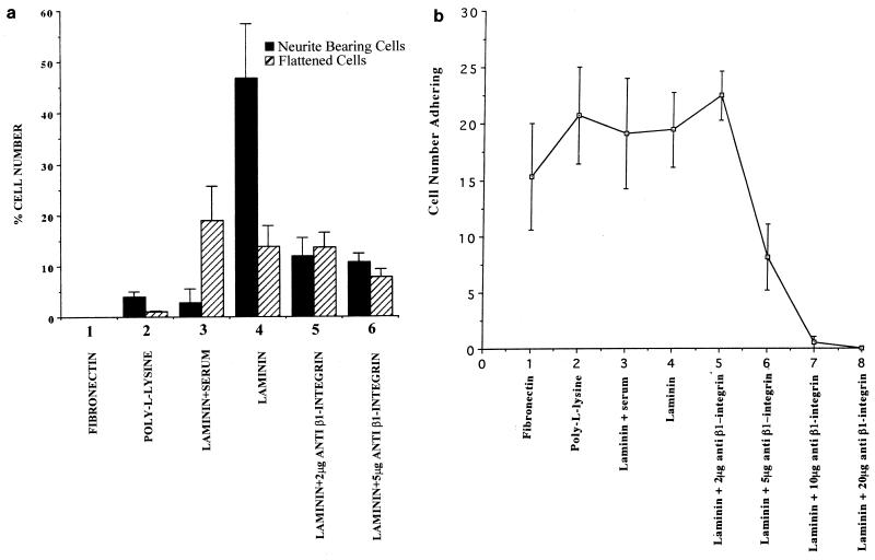
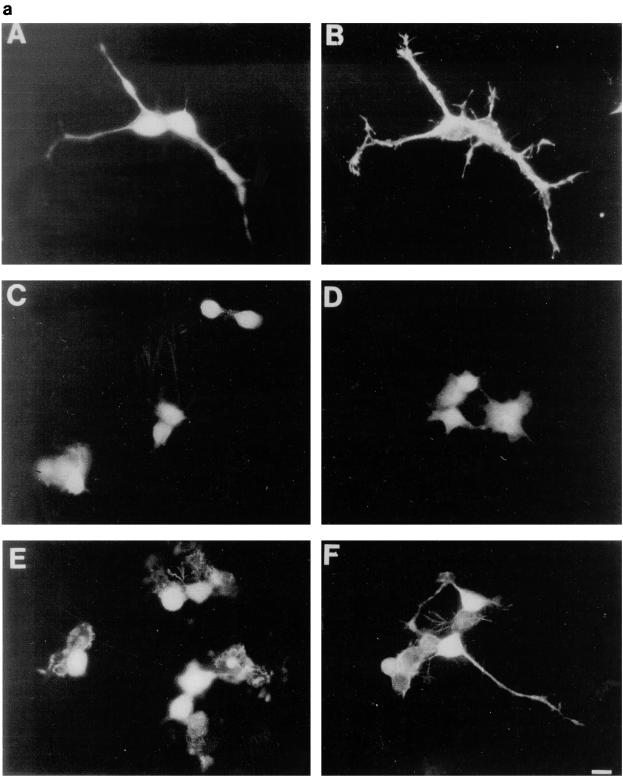
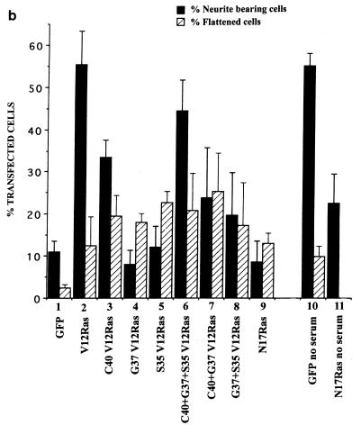
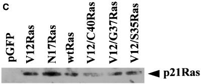
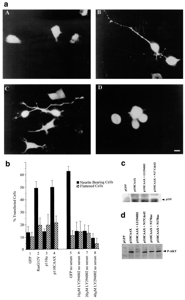
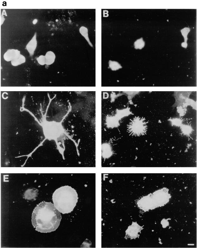
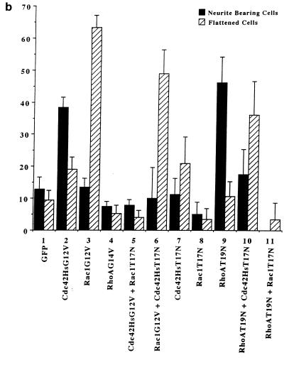
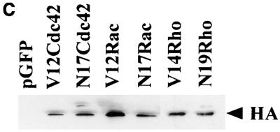
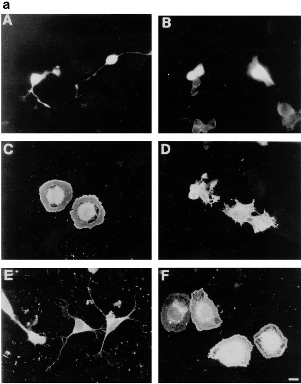
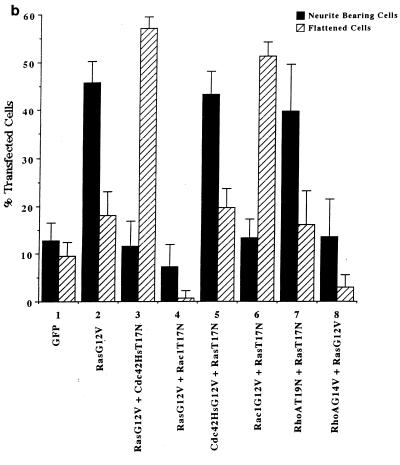
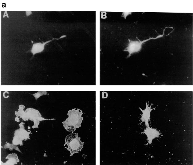
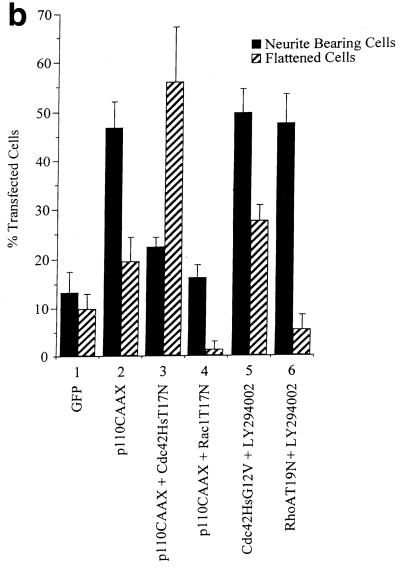
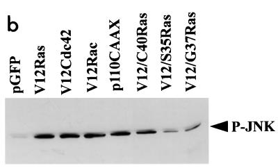
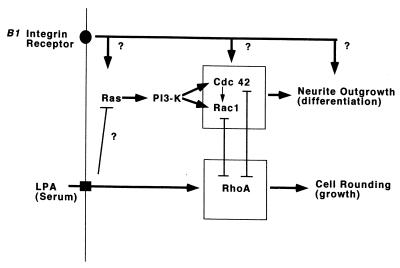
Ras and Rho family GTPases have been ascribed important roles in signalling pathways determining cellular morphology and growth. Here we investigated the roles of the GTPases Ras, Cdc42, Rac1, and Rho and that of phosphatidylinositol 3-kinase (PI 3-kinase) in the pathway leading from serum starvation to neurite outgrowth in N1E-115 neuroblastoma cells. Serum-starved cells grown on a laminin matrix exhibited integrin-dependent neurite outgrowth. Expression of dominant negative mutants of Ras, PI 3-kinase, Cdc42, or Rac1 all blocked this neurite outgrowth, while constitutively activated mutants of Ras, PI 3-kinase, or Cdc42 were each sufficient to promote outgrowth even in the presence of serum. A Ras(H40C;G12V) double mutant which binds preferentially to PI 3-kinase also promoted neurite formation. Activated Ras(G12V)-induced outgrowth required PI 3-kinase activity, but activated PI 3-kinase-induced outgrowth did not require Ras activity. Although activated Rac1 by itself did not induce neurites, neurite outgrowth induced by activated Cdc42(G12V) was Rac1 dependent. Cdc42(G12V)-induced neurites appeared to lose their normal polarization, almost doubling the average number of neurites produced by a single cell. Outgrowth induced by activated Ras or PI 3-kinase required both Cdc42 and Rac1 activity, but Cdc42(G12V)-induced outgrowth did not need Ras or PI 3-kinase activity. Active Rho(G14V) reduced outgrowth promoted by Ras(G12V). Finally, expression of dominant negative Jun N-terminal kinase or extracellular signal-regulated kinase did not inhibit outgrowth, suggesting these pathways are not essential for this process. Our results suggest a hierarchy of signalling where Ras signals through PI 3-kinase to Cdc42 and Rac1 activation (and Rho inactivation), culminating in neurite outgrowth. Thus, in the absence of serum factors, Ras may initiate cell cycle arrest and terminal differentiation in N1E-115 neuroblastoma cells.


Figures

FIG. 1
FIG. 1
Integrin-dependent N1E-115 cell adherence and neurite outgrowth when cells are grown on laminin. (a) The number of cells possessing neurites of at least the length of one cell body or exhibiting a flattened morphology was assessed after plating on the different matrices or following pretreatment with anti-β-1 integrin antibody. Cells were stained with rhodamine-conjugated phalloidin. Numbers are shown as a percentage of each set of 50 cells counted possessing either a neurite or a spread cell body. At least 200 cells were randomly examined, and standard deviations are indicated as vertical bars. (b) The number of cells adhering to a glass chamber slide which had been coated with different extracellular matrices was analyzed by fixing the cells 24 h following plating and staining with rhodamine-conjugated phalloidin. The number of cells per randomly selected field of view was determined on a Zeiss Axioplan fluorescence microscope, and results were compared. At least 20 fields of view were counted for each slide, and the experiments were repeated three times. Cell numbers are shown as the mean number of cells per field of view plus standard deviations shown as vertical bars.
FIG. 2
FIG. 2
Effects of transfection of Ras and Ras mutants on neurite outgrowth. (a) Fluorescence photographs of cells transiently transfected with RasG12V (A and B), wild-type Ras (C), RasT35S;G12V (D), RasE37G;G12V (E), and RasH40C;G12V (F); 20 h following cotransfection with GFP, cells were fixed, stained, and mounted, and the cells expressing GFP were analyzed. Cells were also stained for the neural cell protein neurofilament, shown to be present in the RasG12V-expressing cell (B). (b) Quantification of morphological changes. The morphology of cells expressing GFP was assessed and scored as a percentage of the total number of transfected cells. Cells were designated as neurite bearing if they possessed a neurite at least the length of the cell body and as flattened if they had a large ruffled cell body. Transfections were carried out in the presence of 5% serum except for lanes 8 and 9, in which case transfections were performed in the absence of serum. At least 20 randomly selected fields of view were assessed, and each experiment was repeated at least three times. Standard deviations are shown as vertical bars. (c) Expression of Ras vector constructs in N1E-115 cells. Following transfection, cell lysates were prepared, and equal quantities of proteins were electrophoresed through polyacrylamide gels, Western immunoblotted, and probed with anti-Ras antibodies. wtRas, wild-type Ras.
FIG. 2
FIG. 2
Effects of transfection of Ras and Ras mutants on neurite outgrowth. (a) Fluorescence photographs of cells transiently transfected with RasG12V (A and B), wild-type Ras (C), RasT35S;G12V (D), RasE37G;G12V (E), and RasH40C;G12V (F); 20 h following cotransfection with GFP, cells were fixed, stained, and mounted, and the cells expressing GFP were analyzed. Cells were also stained for the neural cell protein neurofilament, shown to be present in the RasG12V-expressing cell (B). (b) Quantification of morphological changes. The morphology of cells expressing GFP was assessed and scored as a percentage of the total number of transfected cells. Cells were designated as neurite bearing if they possessed a neurite at least the length of the cell body and as flattened if they had a large ruffled cell body. Transfections were carried out in the presence of 5% serum except for lanes 8 and 9, in which case transfections were performed in the absence of serum. At least 20 randomly selected fields of view were assessed, and each experiment was repeated at least three times. Standard deviations are shown as vertical bars. (c) Expression of Ras vector constructs in N1E-115 cells. Following transfection, cell lysates were prepared, and equal quantities of proteins were electrophoresed through polyacrylamide gels, Western immunoblotted, and probed with anti-Ras antibodies. wtRas, wild-type Ras.
FIG. 3
FIG. 3
Involvement of PI 3-kinase in Ras-induced neurite outgrowth. (a) Cells were transiently cotransfected with constructs coding for GFP (A), RasG12V (B), p110CAAX (C), and p110α (D). Slides were fixed and mounted 20 h after transfection, and GFP-expressing cells were analyzed. Note the appearance of the relatively unbranched single neurites produced by RasG12V- and p110CAAX-expressing cells in panels B and C. (b) The morphology of transfected cells was scored as for Fig. 2; cells with a neurite longer than the length of one cell body were scored as neurite bearing, and large ruffled cells were scored as flattened. Numbers were expressed as a percentage of the total number of GFP-expressing cells. Transfection of cells in lanes 1 to 6 was carried out in the presence of serum; transfections in lanes 7 to 11 were performed in the absence of serum. LY294002 was added with the DNA under test, and cells were viewed in a fluorescence microscope. At least 20 randomly selected fields of view were assessed, and each experiment was repeated at least three times. Standard deviations are shown as vertical bars. (c) Expression of p110CAAX vector in N1E-115 cells. Following transfection, cell lysates were prepared, and equal quantities of proteins were electrophoresed on polyacrylamide gels, Western immunoblotted, and probed with anti-p110 antibodies. (d) Activation of Akt in N1E-115 cells by p110CAAX. Following transfection with expression vectors, cell lysates were prepared, and equal quantities of proteins were electrophoresed through polyacrylamide gels, Western immunoblotted, and probed with anti-phospho-Akt antibodies.
FIG. 4
FIG. 4
Cdc42G12V promotes neurite outgrowth dependent on Rac activity. (a) N1E-115 cells were transiently transfected with pGFP and plasmids expressing various HA-tagged p21 GTPase mutants and then stained with anti-HA as follows: (A) rounded GFP-expressing cells; (B) condensed densely stained RhoG14V-expressing cells; (C) Cdc42G12V-expressing cells with many branched neurites, unlike those seen on RasG12V or p110CAAX expression; (D) cells coexpressing Cdc42G12V and Rac1T17N, which are rounded, spiky, and hairy; (E) Rac1G12V-expressing flattened cells with extensive peripheral ruffling and a fried-egg appearance, which was not altered by cotransfection of Cdc42T17N (F). (b) Quantification of morphological changes produced by Rho family GTPase transfection. Cells were stained with anti-HA, and positively stained cells were scored as for Fig. 2 and 3. Expression of GFP and HA correlated in all cases. At least 20 randomly selected fields of view were assessed, and each experiment was repeated at least three times. Standard deviations are shown as vertical bars. (c) Expression of Rho family vector constructs in N1E-115 cells. Following transfection, cell lysates were prepared, and equal quantities of proteins were electrophoresed through polyacrylamide gels, Western immunoblotted, and probed with anti-HA antibodies.
FIG. 4
FIG. 4
Cdc42G12V promotes neurite outgrowth dependent on Rac activity. (a) N1E-115 cells were transiently transfected with pGFP and plasmids expressing various HA-tagged p21 GTPase mutants and then stained with anti-HA as follows: (A) rounded GFP-expressing cells; (B) condensed densely stained RhoG14V-expressing cells; (C) Cdc42G12V-expressing cells with many branched neurites, unlike those seen on RasG12V or p110CAAX expression; (D) cells coexpressing Cdc42G12V and Rac1T17N, which are rounded, spiky, and hairy; (E) Rac1G12V-expressing flattened cells with extensive peripheral ruffling and a fried-egg appearance, which was not altered by cotransfection of Cdc42T17N (F). (b) Quantification of morphological changes produced by Rho family GTPase transfection. Cells were stained with anti-HA, and positively stained cells were scored as for Fig. 2 and 3. Expression of GFP and HA correlated in all cases. At least 20 randomly selected fields of view were assessed, and each experiment was repeated at least three times. Standard deviations are shown as vertical bars. (c) Expression of Rho family vector constructs in N1E-115 cells. Following transfection, cell lysates were prepared, and equal quantities of proteins were electrophoresed through polyacrylamide gels, Western immunoblotted, and probed with anti-HA antibodies.
FIG. 5
FIG. 5
RasG12V-induced neurite outgrowth requires both Cdc42 and Rac1 activity. (a) Neurite outgrowth normally induced by RasG12V transfection (A) is blocked by Cdc42T17N coexpression (C) and Rac1T17N coexpression (D). Cdc42G12V expression still gives neurite outgrowth when cotransfected with RasT17N (E). Rac1G12V cotransfection with RasT17N still gave the typical flattened Rac1-type cells (F) rather than rounded RasT17N cells as shown in panel B. Note the similarity in the appearance of the RasG12V-plus-Cdc42T17N-cotransfected cells (C) to the fried-egg cells seen following Rac1G12V transfection (Fig. 4a, panel E) and the hairy cells produced with RasG12V plus Rac1T17N coexpression, akin to the Cdc42G12V-plus-Rac1T17N-coexpressing cells shown previously (Fig. 4a, panel D). (b) Quantification of the above morphologies. Cells coexpressing GFP or stained with HA were scored as being neurite bearing, flattened, or rounded, and this was expressed as a percentage of the total number of positively expressing cells in a field of view. At least 20 randomly selected fields of view were assessed, and each experiment was repeated at least three times. Standard deviations are shown as vertical bars.
FIG. 5
FIG. 5
RasG12V-induced neurite outgrowth requires both Cdc42 and Rac1 activity. (a) Neurite outgrowth normally induced by RasG12V transfection (A) is blocked by Cdc42T17N coexpression (C) and Rac1T17N coexpression (D). Cdc42G12V expression still gives neurite outgrowth when cotransfected with RasT17N (E). Rac1G12V cotransfection with RasT17N still gave the typical flattened Rac1-type cells (F) rather than rounded RasT17N cells as shown in panel B. Note the similarity in the appearance of the RasG12V-plus-Cdc42T17N-cotransfected cells (C) to the fried-egg cells seen following Rac1G12V transfection (Fig. 4a, panel E) and the hairy cells produced with RasG12V plus Rac1T17N coexpression, akin to the Cdc42G12V-plus-Rac1T17N-coexpressing cells shown previously (Fig. 4a, panel D). (b) Quantification of the above morphologies. Cells coexpressing GFP or stained with HA were scored as being neurite bearing, flattened, or rounded, and this was expressed as a percentage of the total number of positively expressing cells in a field of view. At least 20 randomly selected fields of view were assessed, and each experiment was repeated at least three times. Standard deviations are shown as vertical bars.
FIG. 6
FIG. 6
p110CAAX-induced neurite outgrowth requires Cdc42Hs and Rac1 activity. (a) At 20 h after transfection, p110CAAX expression produced neurites (A) which also stained positively for neurofilament protein (B). Transient cotransfection of p110CAAX and Cdc42T17N gave flattened Rac1-type cells and no neurite outgrowth (C). Coexpression of p110CAAX and dominant negative Rac1T17N also inhibited PI 3-kinase-induced outgrowth, but cells exhibited the hairy cell morphology (D). (b) Quantification of transfected cell morphologies was carried out as described in legend to Fig. 5.
FIG. 6
FIG. 6
p110CAAX-induced neurite outgrowth requires Cdc42Hs and Rac1 activity. (a) At 20 h after transfection, p110CAAX expression produced neurites (A) which also stained positively for neurofilament protein (B). Transient cotransfection of p110CAAX and Cdc42T17N gave flattened Rac1-type cells and no neurite outgrowth (C). Coexpression of p110CAAX and dominant negative Rac1T17N also inhibited PI 3-kinase-induced outgrowth, but cells exhibited the hairy cell morphology (D). (b) Quantification of transfected cell morphologies was carried out as described in legend to Fig. 5.
FIG. 7
FIG. 7
Neurite outgrowth in N1E-115 cells does not require the JNK or ERK pathway. (a) Transfection of SEK-AL (A) or MEK-AL (E) dominant negative constructs in serum-starved conditions and cotransfections as indicated. At 20 h after transfection, cells were fixed and stained. (b) Activation of JNK in N1E-115 cells following transfection with expression vectors. Following transfection, cell lysates were prepared, and equal quantities of proteins were electrophoresed through polyacrylamide gels, Western immunoblotted, and probed with anti-phospho-JNK antibodies.
FIG. 7
FIG. 7
Neurite outgrowth in N1E-115 cells does not require the JNK or ERK pathway. (a) Transfection of SEK-AL (A) or MEK-AL (E) dominant negative constructs in serum-starved conditions and cotransfections as indicated. At 20 h after transfection, cells were fixed and stained. (b) Activation of JNK in N1E-115 cells following transfection with expression vectors. Following transfection, cell lysates were prepared, and equal quantities of proteins were electrophoresed through polyacrylamide gels, Western immunoblotted, and probed with anti-phospho-JNK antibodies.
FIG. 8
FIG. 8
Neurite outgrowth following serum deprivation signals through Ras, PI 3-kinase, and Cdc42 and Rac1. Activation of Ras, PI 3-kinase or Cdc42 (but not activation of Rac1 alone) is able to promote neurite outgrowth even in the presence of serum factors, but in each case it requires the presence of a laminin matrix which signals via integrin receptors. It is not known whether the GTPases signal upstream or downstream from the integrins or whether the integrin requirement is independent of these GTPases. Inhibition of Rho also results in neurite outgrowth, and Rho has been shown to compete with Cdc42 and Rac1 (32, 63). The mechanism responsible for the activation of Cdc42 and Rac1 by PI 3-kinase is not clear but probably involves phosphatidylinositol 3,4,5-triphosphate-dependent increased nucleotide exchange activity and/or inhibition of GTPase activity for the p21s (13).

Similar articles

Cited by

References

    1. Barbacid M. Ras genes. Annu Rev Biochem. 1987;56:779–827. - PubMed
    1. Bar-Sagi D, Feramisco J R. Microinjection of the ras oncoprotein into PC12 cells induces morphological differentiation. Cell. 1985;42:841–848. - PubMed
    1. Bar-Sagi D, Feramisco J R. Induction of membrane ruffling and fluid-phase pinocytosis in quiescent fibroblasts by ras proteins. Science. 1986;233:1061–1068. - PubMed
    1. Bollag G, McCormick F. Regulators and effectors of Ras proteins. Annu Rev Cell Biol. 1991;7:601–632. - PubMed
    1. Boguski M S, McCormick F. Proteins regulating Ras and its relatives. Nature. 1993;366:643–654. - PubMed

Publication types