. 2000 Jan;20(1):261-72.
doi: 10.1128/MCB.20.1.261-272.2000.

Establishment of distinct MyoD, E2A, and twist DNA binding specificities by different basic region-DNA conformations

Affiliations

Establishment of distinct MyoD, E2A, and twist DNA binding specificities by different basic region-DNA conformations

T Kophengnavong et al. Mol Cell Biol. 2000 Jan.

Abstract

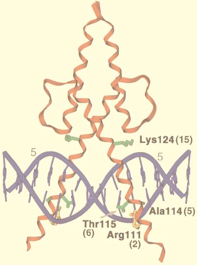
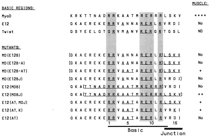
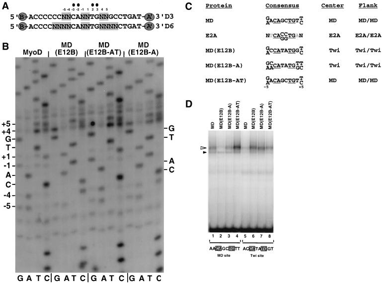
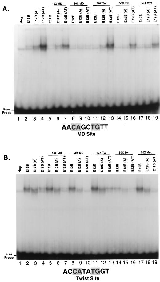
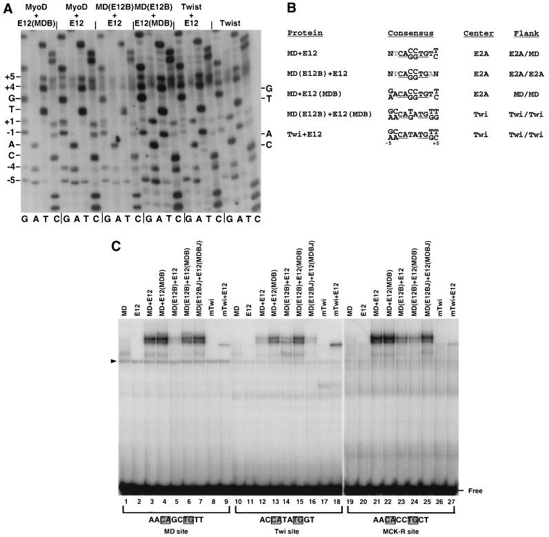
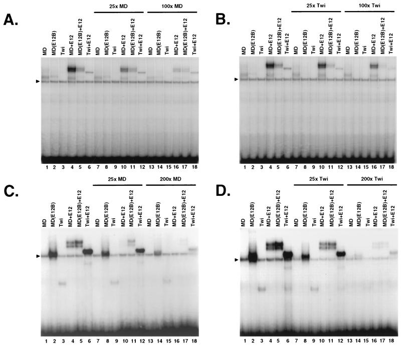
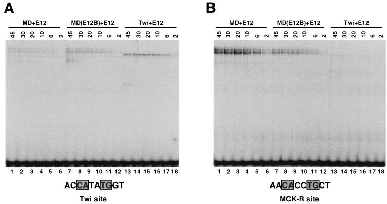
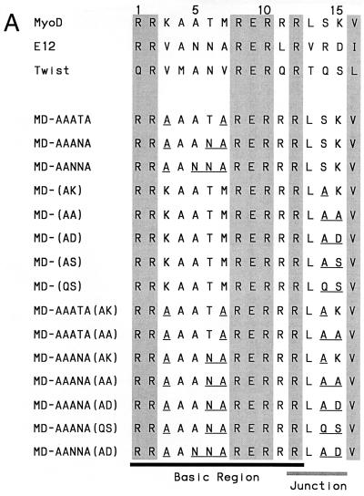
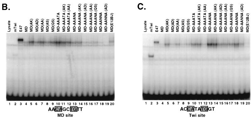
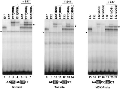
Basic helix-loop-helix (bHLH) proteins perform a wide variety of biological functions. Most bHLH proteins recognize the consensus DNA sequence CAN NTG (the E-box consensus sequence is underlined) but acquire further functional specificity by preferring distinct internal and flanking bases. In addition, induction of myogenesis by MyoD-related bHLH proteins depends on myogenic basic region (BR) and BR-HLH junction residues that are not essential for binding to a muscle-specific site, implying that their BRs may be involved in other critical interactions. We have investigated whether the myogenic residues influence DNA sequence recognition and how MyoD, Twist, and their E2A partner proteins prefer distinct CAN NTG sites. In MyoD, the myogenic BR residues establish specificity for particular CAN NTG sites indirectly, by influencing the conformation through which the BR helix binds DNA. An analysis of DNA binding by BR and junction mutants suggests that an appropriate BR-DNA conformation is necessary but not sufficient for myogenesis, supporting the model that additional interactions with this region are important. The sequence specificities of E2A and Twist proteins require the corresponding BR residues. In addition, mechanisms that position the BR allow E2A to prefer distinct half-sites as a heterodimer with MyoD or Twist, indicating that the E2A BR can be directed toward different targets by dimerization with different partners. Our findings indicate that E2A and its partner bHLH proteins bind to CAN NTG sites by adopting particular preferred BR-DNA conformations, from which they derive differences in sequence recognition that can be important for functional specificity.


Figures

FIG. 1
FIG. 1
A MyoD-DNA complex. In this X-ray crystallographic structure (35), a MyoD homodimer is bound to the sequence AACAGCTGTT, which corresponds to its preferred recognition consensus (9). Residues are numbered as in full-length MyoD, and their positions as specified in Fig. 2 and the text are indicated in parentheses. Binding site positions ±5 (numbered as in Fig. 3A) are indicated by grey numerals. Side chains are shown only for the myogenic residues (green) (18) and Arg 111 (R2) (gold).
FIG. 2
FIG. 2
Myogenic activity of MyoD and E12 BR and junction mutants. Each of these mutants has been described previously (18, 57), and their sequences are compared with sequences from mouse MyoD, E12, and Twist. Amino acids that are identical to those of MyoD are underlined, positions that are conserved in most bHLH proteins are shaded, and entire BR and junction regions that have been swapped are bracketed. The column at the right indicates the relative activities of these proteins when assayed previously by transfection for conversion of cultured cells into muscle (18, 57); activity is denoted as ++++ (frequency of myogenic conversion obtained with wild-type MyoD), ++ (30 to 50% of that obtained with MyoD), + (5 to 30% of that obtained with wild-type MyoD), No (myogenic conversion was not detected), or ND (not done).
FIG. 3
FIG. 3
In vitro selection assay of binding site preferences. (A) Core sequences of the random sequence oligonucleotide libraries D3 and D6 (8, 9). In each library, the bases shown are flanked by sequences which correspond to primers (A and B) that allow selected sequences to be recovered by PCR. A′ indicates that primer A corresponds to the opposite strand. (B) Sequences of preferred binding sites. Starting with the D6 oligonucleotide random sequence library (A), three rounds of sequential selection and PCR amplification were performed for binding to the proteins indicated. A sample of the final selected population of binding sites was then sequenced directly as a pool and analyzed by autoradiography. The MyoD preferences at positions ±1 described previously (9) are more prominent after additional selection rounds (not shown). (C) Summary of sequence preferences identified by in vitro selection in panel B. MyoD and E2A homodimer preferences were described in reference . Binding site positions are numbered as in panel B, and grey letters indicate bases that were selected against. The CAN NTG consensus that was fixed in these experiments is underlined. Twi, Twist. (D) Binding of MyoD BR mutants to individual oligonucleotide sites, which differed only at the sequences shown. In this EMSA, which was analyzed by phosphorimaging, each sample contained the indicated in vitro-translated protein at a concentration of 40 pM and DNA that was labeled to the same specific activity at 550 pM. Specific and background species are indicated by open and closed triangles, respectively.
FIG. 4
FIG. 4
Specificity of MyoD BR mutant DNA binding. (A) Competition analysis of binding to the labeled MyoD preferred site, analyzed by EMSA and autoradiography. The indicated in vitro-translated proteins and DNA labeled to the same specific activity were present at concentrations of 50 and 900 pM, respectively. When the samples were mixed, unlabeled competitor DNA sites were added at the indicated ratios relative to the labeled probe. Tw, Twist. (B) Competition analysis of binding to the Twist preferred site, performed as for panel A.
FIG. 5
FIG. 5
Binding site preferences of MyoD, E2A, and Twist heterodimer complexes. (A) In vitro selection analysis of binding site preferences. Four rounds of selection from the D3 library (Fig. 3A) were performed as for each in vitro-translated protein complex. In each case, the heterodimer complex could be easily identified in the EMSA on the basis of mobility (9), particularly because E12 homodimers bind DNA poorly (Fig. 9). In the Twist homodimer selection, binding to Twist-E12 heterodimers was selected for in the first round, because of the relatively low level of Twist homodimer binding. Subsequent rounds were performed with Twist homodimers. Each sample was analyzed by sequencing and autoradiography as for Fig. 3B. (B) Summary of sequence preferences identified in panel A, depicted as in Fig. 3C. MyoD-E2A heterodimer preferences were also described previously (9). Twi, Twist. (C) Binding of bHLH heterodimers to individual preferred sites, analyzed by EMSA and phosphorimaging. E2A-derived proteins were present at a concentration of 8 pM, and Twist and MyoD-derived proteins were present at 19 pM. The indicated DNA sites that had been labeled to the same specific activity were present at 550 pM. The MCK-R site differs from the others only at the positions shown. A background species is indicated by a triangle.
FIG. 6
FIG. 6
Binding competition analysis of DNA binding by bHLH heterodimers. (A and B) Binding of the indicated protein complexes to the labeled MyoD site (Fig. 3D) was competed by addition of an unlabeled binding site at ratios indicated above the gel. These experiments were performed and analyzed as for Fig. 4 except that labeled DNA was present at 600 pM, E12 protein present at 8 pM, and all other proteins were present at 19 pM. Twi, Twist. (C and D) Binding of the indicated protein complexes to the labeled Twist site (Fig. 3D) was competed by addition of the indicated unlabeled sites. These experiments were performed described for panel A and B except that labeled DNA was present at 1.1 nM, and they were analyzed by autoradiography. Note that the gel shown in panel C was exposed longer than that shown in panel D, as indicated by comparison of lanes 1 to 6. A background species is indicated by a triangle.
FIG. 7
FIG. 7
Protein titration of DNA binding by bHLH heterodimers. (A) Binding to the Twist (Twi) site, analyzed by EMSA and phosphorimaging. In each experiment, E12 was present at 8 pM and DNA that had been labeled to the same specific activity was present at 5 pM. The indicated partner proteins were present at the concentrations (picomolar) shown above the gel. (B) Binding to the MCK-R site, analyzed as for panel A.
FIG. 8
FIG. 8
Effects of bHLH BR and BR-HLH junction residues on MyoD binding preferences. (A) Mutagenesis analysis of the MyoD BR and junction. MyoD BR mutant sequences are compared with the MyoD, E12, and Twist BR sequences (Fig. 2). Conserved bHLH residues are shaded, and residues that are altered within full-length MyoD are underlined. (B) Binding of MyoD mutants described in panel A to the MyoD preferred site. These mutants are compared with the indicated wild-type proteins, and binding is assayed as for Fig. 3D except that each protein is present at 40 pM and DNA labeled to the same specific activity is present at 400 pM. E47 is an alternatively spliced E2A protein that binds DNA well as a homodimer (40). Twi, Twist. (C) Binding of MyoD mutants to the Twist preferred site, assayed as for panel B.
FIG. 8
FIG. 8
Effects of bHLH BR and BR-HLH junction residues on MyoD binding preferences. (A) Mutagenesis analysis of the MyoD BR and junction. MyoD BR mutant sequences are compared with the MyoD, E12, and Twist BR sequences (Fig. 2). Conserved bHLH residues are shaded, and residues that are altered within full-length MyoD are underlined. (B) Binding of MyoD mutants described in panel A to the MyoD preferred site. These mutants are compared with the indicated wild-type proteins, and binding is assayed as for Fig. 3D except that each protein is present at 40 pM and DNA labeled to the same specific activity is present at 400 pM. E47 is an alternatively spliced E2A protein that binds DNA well as a homodimer (40). Twi, Twist. (C) Binding of MyoD mutants to the Twist preferred site, assayed as for panel B.
FIG. 9
FIG. 9
DNA binding by E12 mutants. DNA binding by the indicated protein complexes is assayed as for Fig. 5C except that all E12 derivatives are present at 8 pM and E47 is present at 19 pM. A protein-DNA complex of intermediate mobility that corresponds to E47-E12 heterodimers is indicated by an asterisk, and a background species is indicated by a closed triangle.

Similar articles

Cited by

References

    1. Aksan I, Goding C R. Targeting the microphthalmia basic helix-loop-helix-leucine zipper transcription factor to a subset of E-box elements in vitro and in vivo. Mol Cell Biol. 1998;18:6930–6938. - PMC - PubMed
    1. Anthony-Cahill S J, Benfield P A, Fairman R, Wasserman Z R, Brenner S L, Stafford W F, Altenbach C, Hubbell W L, DeGrado W F. Molecular characterization of helix-loop-helix peptides. Science. 1992;255:979–983. - PubMed
    1. Bengal E, Flores O, Rangarajan P N, Chen A, Weintraub H, Verma I M. Positive control mutations in the MyoD basic region fail to show cooperative DNA binding and transcriptional activation in vitro. Proc Natl Acad Sci USA. 1994;91:6221–6225. - PMC - PubMed
    1. Biesiada E, Hamamori Y, Kedes L, Sartorelli V. Myogenic basic helix-loop-helix proteins and Sp1 interact as components of a multiprotein transcriptional complex required for activity of the human cardiac alpha-actin promoter. Mol Cell Biol. 1999;19:2577–2584. - PMC - PubMed
    1. Black B L, Molkentin J D, Olson E N. Multiple roles for the MyoD basic region in transmission of transcriptional activation signals and interaction with MEF2. Mol Cell Biol. 1998;18:69–77. - PMC - PubMed

Publication types

LinkOut - more resources