. 2000 Jan;20(1):273-85.
doi: 10.1128/MCB.20.1.273-285.2000.

Regulation of a senescence checkpoint response by the E2F1 transcription factor and p14(ARF) tumor suppressor

Affiliations

Regulation of a senescence checkpoint response by the E2F1 transcription factor and p14(ARF) tumor suppressor

G P Dimri et al. Mol Cell Biol. 2000 Jan.

Abstract

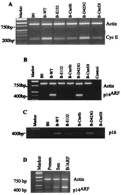
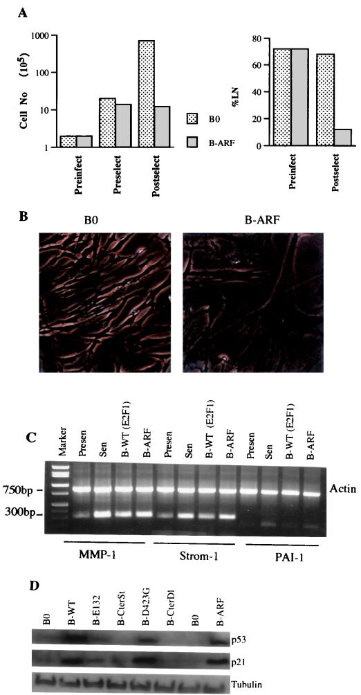
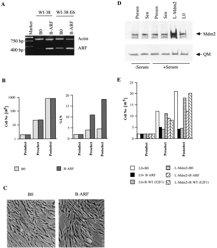
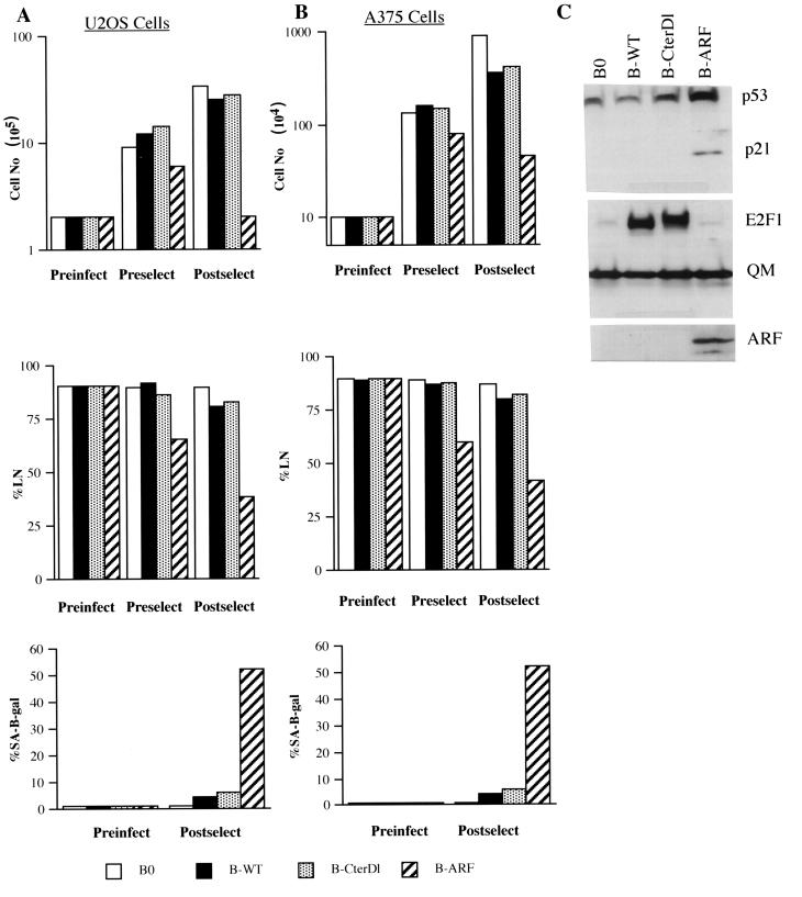
Normal cells do not divide indefinitely due to a process known as replicative senescence. Human cells arrest growth with a senescent phenotype when they acquire one or more critically short telomeres as a consequence of cell division. Recent evidence suggests that certain types of DNA damage, chromatin remodeling, and oncogenic forms of Ras or Raf can also elicit a senescence response. We show here that E2F1, a multifunctional transcription factor that binds the retinoblastoma (pRb) tumor suppressor and that can either promote or suppress tumorigenesis, induces a senescent phenotype when overexpressed in normal human fibroblasts. Normal human cells stably arrested proliferation and expressed several markers of replicative senescence in response to E2F1. This activity of E2F1 was independent of its pRb binding activity but dependent on its ability to stimulate gene expression. The E2F1 target gene critical for the senescence response appeared to be the p14(ARF) tumor suppressor. Replicatively senescent human fibroblasts overexpressed p14(ARF), and ectopic expression of p14(ARF) in presenescent cells induced a phenotype similar to that induced by E2F1. Consistent with a critical role for p14(ARF), cells with compromised p53 function were immune to senescence induction by E2F1, as were cells deficient in p14(ARF). Our findings support the idea that the senescence response is a critical tumor-suppressive mechanism, provide an explanation for the apparently paradoxical roles of E2F1 in oncogenesis, and identify p14(ARF) as a potentially important mediator of the senescent phenotype.


Figures

FIG. 1
FIG. 1
Analysis of E2F1 proteins used in this study. (A) Diagram of wild-type (WT) and mutant (E132, CterSt, D423G, CterDl) E2F1 proteins described in the text. (B) Expression levels. Normal (WI-38) or E6-expressing (WI-38-E6) human fibroblasts were infected with control (B0) or E2F1-expressing retroviruses (B-WT, B-E132, B-CterSt, B-D423G, B-CterDl). Protein lysates were prepared one passage after antibiotic selection and analyzed by Western blotting for the levels of E2F1 and QM (control) (18) proteins. The presence of two E2F1 protein species, migrating with slightly different mobilities (more prominent in E6-expressing cells), is consistent with phosphorylated and unphosphorylated forms. Under our SDS-PAGE conditions, wild-type and mutant E2F1 proteins migrated with approximately the same mobilities, despite slight differences in size. The estimated sizes of the E2F1 proteins are 52 kDa for WT, E132, and D423G; 46 kDa for CterSt; and 59 kDa for CterDl (due to the addition of 58 C-terminal amino acids from the vector). (C) pRb-binding activities. WI-38-E6 cells were infected with control or E2F1-expressing retroviruses, made quiescent by serum deprivation (to reduce endogenous E2F1), and lysed. Then, 500 μg of protein lysate was immunoprecipitated with anti-E2F1 antibody and analyzed for pRb by Western blotting (IP/Western). In parallel, 15 μg of protein was analyzed without immunoprecipitation (Western) for pRb and α-tubulin (control). (D) Transactivation activities. E6-expressing cells infected with control or E2F1-expressing viruses were cotransfected with a normalization vector and either pGL2, a promoterless luciferase vector, or E2F-luc, in which luciferase is driven by E2F binding sites (42); extracts were prepared and analyzed as described in Materials and Methods. The results shown are the amounts of normalized E2F-luc activity relative to the amount of normalized pGL2 activity.
FIG. 2
FIG. 2
Effects of E2F1 proteins on cell proliferation, DNA synthesis, and apoptosis. (A) Proliferation of normal human cells. WI-38 normal human fibroblasts (Preinfect; 2 × 10⁵ cells) were mock infected (Mock) or infected with control (B0) or E2F1-expressing (wild-type [WT], E132, CterSt, D423G, CterDl) retroviruses. Three days after infection, cell number was determined (Preselect). Infected cells were then selected with puromycin for 5 days and cultured in medium without antibiotic for an additional 3 days; mock-infected cells were cultured without antibiotic for 8 days. The cell number was then again determined (Postselect). Similar results were obtained in at least three independent experiments using different batches of virus. (B) DNA synthesis in normal human cells. WI-38 cells (Preinfect) were infected and selected as described for panel A. After selection, [3H]thymidine was added in antibiotic-free medium and the cells were processed for autoradiography 3 days later (Postselect). A minimum of 200 cells were counted to determine the percentages of radiolabeled nuclei (%LN). (C) Proliferation of E6-expressing human cells. WI-38 cells were infected with an LXSN retrovirus carrying E6 and selected in G-418, as described in Materials and Methods (WI-38-E6 cells). The resulting WI-38-E6 cultures were then infected with E2F1-expressing retroviruses, selected in puromycin, and counted as described for normal WI-38 cells (A). (D) Apoptosis in normal human and immortal mouse cells. WI-38 and mouse NIH 3T3 fibroblasts were infected with control (B0) or E2F1-expressing (B-WT) retroviruses, as described in Materials and Methods. Seventy-two hours later, detached (floating) cells were collected by gentle centrifugation. DNA was isolated from the floating cells and the remaining attached cells and analyzed as described in Materials and Methods. WI-38 cells also showed no DNA fragmentation if they were selected in puromycin prior to DNA isolation (not shown). Left lane, DNA size markers. (E) Apoptosis in normal human cells. WI-38 cells were infected with control (B0) or E2F1-expressing (B-WT) retroviruses, selected in puromycin for 3 days, and plated on chambered glass slides; 24 h later, they were assayed in situ for DNA fragmentation by TUNEL, and then stained with DAPI (4′,6′-diamidino-α-phenylindole) as described in Materials and Methods. HT1080 cells treated for 16 h with 10 μM MG132, a proteasome inhibitor that induces apoptosis in human tumor cells (37), served as a positive control for the reaction. No TUNEL-positive cells were observed among >1,000 B0- or B-WT-infected WI-38 cells (<0.1% apoptosis), whereas 52 of 611 TUNEL-positive cells were observed among MG132-treated HT1080 cells (8.5% apoptosis). (F) Stability of E2F1 growth arrest. WI-38 cells were infected and selected as described for panel A. After selection 106 cells were plated in antibiotic-free medium (day 1). The numbers of cells were determined 2 (day 3) and 9 days (day 10) later.
FIG. 3
FIG. 3
E2F1 proteins induce a senescence-like phenotype. (A) Morphology and SA-β-Gal expression. WI-38 cells were infected with the indicated E2F1-expressing retroviruses, selected, and, 2 to 3 days thereafter, stained for SA-β-Gal, as described in Materials and Methods. The cells were photographed under phase-contrast optics. WT, wild type. (B) Expression of matrix-remodeling genes. RNA, isolated from WI-38 cells infected as described for panel A, was analyzed for interstitial collagenase (MMP-1), stromelysin-1 (Strom-1), PAI-1, and β-actin (control) mRNA by RT-PCR, as described in Materials and Methods. Signals were normalized to β-actin, and the MMP-1, stromelysin-1, and PAI-1 mRNA levels in control cells (B0) were each set at 1. Relative to that by B0, levels of induction by B-WT were 2.7, 2.2, and 1.8, respectively; levels of induction by B-E132 were 0.9, 1.0, and 1.1, respectively; and levels of induction by B-CterDl were 0.9, 1.2, and 1.0, respectively.
FIG. 4
FIG. 4
Induction of cyclin E, p14ARF, and p16INK4a by E2F1 proteins. (A) Cyclin E. RNA was isolated from postselected WI-38 cells that had been infected with the indicated E2F1-expressing retroviruses (WT, wild type) and analyzed by semiquantitative RT-PCR for β-actin (control) and cyclin E mRNA. The cyclin E signal was normalized to β-actin, and the expression level in control infected cells (B0) was set at 1. Relative to that for B0, levels of induction of cyclin E were as follows: B-WT, 3.2; B-E132, 1.2; B-CterSt, 1.3; B-D423G, 3.1; B-CterSt, 1.2. (B) p14ARF. RNA was isolated, analyzed for p14ARF and β-actin, and normalized as described for panel A. Relative to that for B0, the levels of induction of p14ARF were as follows: B-WT, 5.7; B-E132, 1.6; B-CterSt, 0.9; B-D423G, 5.5; B-CterSt, 1.0. (C) p16INK4a. RNA was isolated, analyzed for p16INK4a and β-actin, and normalized as described for panel A. p16INK4a was analyzed in the same experiment as p14ARF and has the same β-actin (B) control. (D) Expression of p14ARF in presenescent, senescent, and retrovirally infected cells. RNA was isolated from presenescent (Presen) and senescent (Sen) WI-38 cells, as well as WI-38 cells infected with the p14ARF retrovirus, and cells were cultured in serum-containing medium. RNA was analyzed and normalized as described for panel A, and the p14ARF expression in senescent cells was set at 1. Relative to that for senescent cells, p14ARF expression levels were 0.15 in presenescent cells and 3 in retrovirally infected cells.
FIG. 5
FIG. 5
Senescent phenotype induced by p14ARF. (A) Growth inhibition. Cell number was determined prior to infection by control (B0) or p14ARF-expressing (B-ARF) retroviruses (Preinfect), prior to selection (Preselect), and after selection (Postselect), as described in the legend to Fig. 2A. Postselected cells were also given [3H]thymidine for 72 h to determine the fraction of cells that were capable of synthesizing DNA (%LN). (B) Morphology and SA-β-Gal expression. Postselected cells were stained for SA-β-Gal and photographed under phase-contrast optics. (C) Expression of matrix-remodeling genes. RNA was isolated from postselected WI-38 cells, as well as presenescent and senescent WI-38 cells and analyzed by semiquantitative RT-PCR for actin (control) mRNA and interstitial collagenase (MMP-1), stromelysin-1 (Strom-1), and PAI-1 mRNA. See Table 2 for quantitation. (D) Induction of p53 and p21 by 14ARF and E2F1. Protein extracts were prepared from postselected cells and analyzed by Western blotting for p53, p21, and tubulin (control).
FIG. 6
FIG. 6
Senescent phenotype induced by E2F1 and p14ARF is p53 dependent. (A) p14ARF expression in normal and E6-expressing cells. WI-38-E6 cells were infected with control (B0) or p14ARF-expressing (B-ARF) retroviruses and selected. RNA was isolated from postselected cells and analyzed by RT-PCR for actin (control) and p14ARF mRNA. The same analysis performed on normal WI-38 cells is shown for comparison. (B) Growth inhibition by p14ARF in E6-expressing cells. WI-38-E6 cells were infected and selected as described for panel A. Cell numbers were determined prior to infection (Preinfect) and selection (Preselect) and after selection (Postselect). Postselected cells were labeled with [3H]thymidine for 72 h. A minimum of 200 cells were counted to determine the percentage of radiolabeled nuclei (%LN). (C) Morphology and SA-β-Gal expression. Postselected WI-38-E6 cells were stained for SA-β-Gal and photographed under phase-contrast optics. (D) Mdm2 expression. Protein extracts were prepared from presenescent and senescent WI-38 cells maintained for 3 days in 0.2 (−Serum) or 10% (+Serum) serum, and proliferating cells were infected with control (B0) or Mdm2 (B-Mdm2)-expressing retroviruses. Extracts were analyzed by Western blotting for Mdm2 and QM (control) proteins. (E) Growth inhibition in Mdm2-expressing cells. WI-38 cells were infected with control (L0) or Mdm2-expressing (L-Mdm2) viruses, selected in G-418, and superinfected with control (B0), p14ARF (B-ARF), or wild-type E2F1 [WT (E2F1)] viruses, and selected in puromycin. Cells were counted prior to infection (Preinfect) and selection (Preselect) and 3 days after selection (Postselect).
FIG. 7
FIG. 7
Senescent phenotype induced by E2F1 is p14ARF dependent. (A) U2OS cells were infected with control (B0)-, wild-type E2F1 (B-WT)-, transactivation defective CterD1 E2F1 (B-CterD1)-, or p14ARF (B-ARF)-expressing retroviruses and selected in puromycin. The cells were counted or labeled with [3H]thymidine for 3 days to determine the percentage that synthesized DNA (%LN) prior to infection (Preinfect), prior to selection (Preselect), or 2 to 3 days after selection (Postselect). In parallel, the cells were stained for SA-β-Gal activity prior to infection (Preinfect) and after selection (Postselect). For each determination of percent labeled nuclei and SA-β-Gal activity, at least 400 cells were counted from two independent culture dishes. (B) A375 cells were infected and analyzed for cell number, percent labeled nuclei, and SA-β-Gal activity, as described for U2OS cells (A). (C) Protein lysates were prepared from postselected U2OS cells infected with control (B0)-, wild-type E2F1 (B-WT)-, CterDl E2F1 (B-CterDl)-, or p14ARF (B-ARF)-expressing retroviruses and analyzed by Western blotting for p53, p21, E2F1, p14ARF, and QM (control) protein.

Similar articles

Cited by

References

    1. Afshari C A, Vojta P J, Annab L A, Futreal P A, Willard T B, Barrett J C. Investigation of the role of G1/S cell cycle mediators in cellular senescence. Exp Cell Res. 1993;209:231–237. - PubMed
    1. Alcorta D A, Xiong Y, Phelps D, Hannon G, Beach D, Barrett J C. Involvement of the cyclin-dependent kinase inhibitor p16 (INK4a) in replicative senescence of normal human fibroblasts. Proc Natl Acad Sci USA. 1996;93:13742–13747. - PMC - PubMed
    1. Atadja P, Wong H, Garkavstev I, Veillette C, Riabowol K. Increased activity of p53 in senescing fibroblasts. Proc Natl Acad Sci USA. 1995;92:8348–8352. - PMC - PubMed
    1. Bates S, Phillips A C, Clark P A, Stott F, Peters G, Ludwig R L, Voudsen K H. p14ARF links the tumor suppressors RB and p53. Nature. 1998;395:124–125. - PubMed
    1. Blattner C, Sparks A, Lane D. Transcription factor E2F-1 is upregulated in response to DNA damage in a manner analogous to that of p53. Mol Cell Biol. 1999;19:3704–3713. - PMC - PubMed

Publication types