. 2000 Jan;20(1):402-15.
doi: 10.1128/MCB.20.1.402-415.2000.

Redox-regulated recruitment of the transcriptional coactivators CREB-binding protein and SRC-1 to hypoxia-inducible factor 1alpha

Affiliations

Redox-regulated recruitment of the transcriptional coactivators CREB-binding protein and SRC-1 to hypoxia-inducible factor 1alpha

P Carrero et al. Mol Cell Biol. 2000 Jan.

Abstract

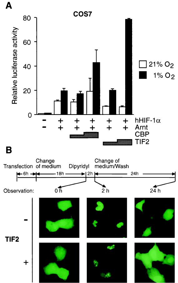
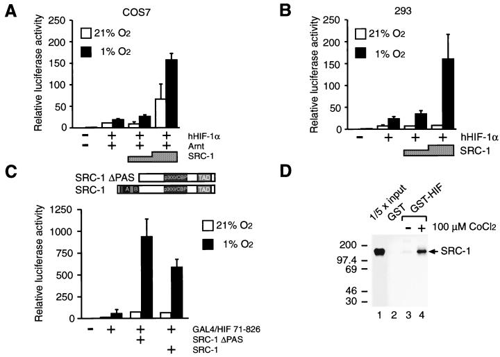
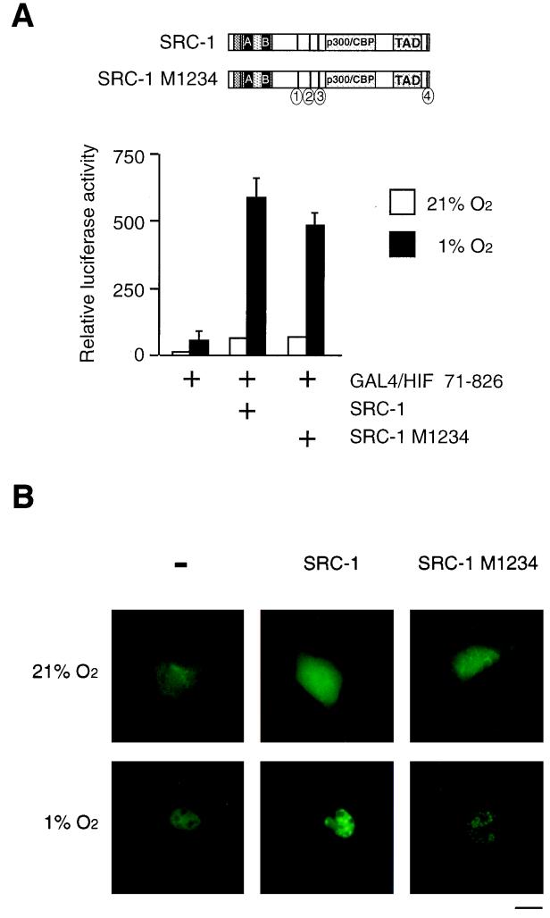
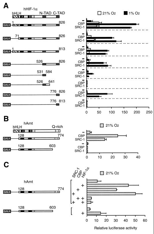
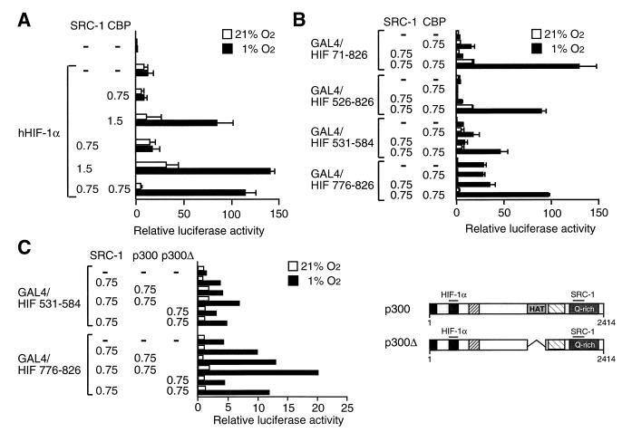
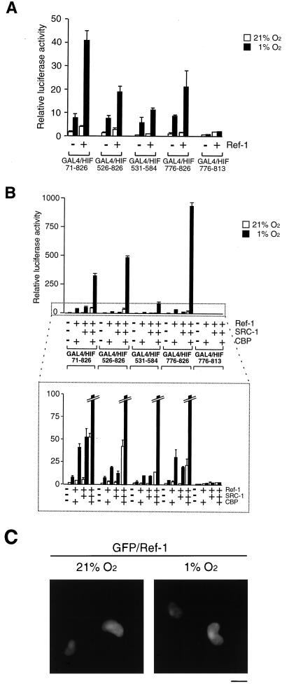
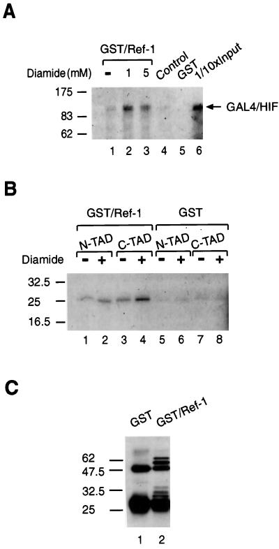
Hypoxia-inducible factor 1alpha (HIF-1alpha) functions as a transcription factor that is activated by decreased cellular oxygen concentrations to induce expression of a network of genes involved in angiogenesis, erythropoiesis, and glucose homeostasis. Here we demonstrate that two members of the SRC-1/p160 family of transcriptional coactivators harboring histone acetyltransferase activity, SRC-1 and transcription intermediary factor 2 (TIF2), are able to interact with HIF-1alpha and enhance its transactivation potential in a hypoxia-dependent manner. HIF-1alpha contains within its C terminus two transactivation domains. The hypoxia-inducible activity of both these domains was enhanced by either SRC-1 or the CREB-binding protein (CBP)/p300 coactivator. Moreover, at limiting concentrations, SRC-1 produced this effect in synergy with CBP. Interestingly, this effect was strongly potentiated by the redox regulatory protein Ref-1, a dual-function protein harboring DNA repair endonuclease and cysteine reducing activities. These data indicate that all three proteins, CBP, SRC-1, and Ref-1, are important components of the hypoxia signaling pathway and have a common function in regulation of HIF-1alpha function in hypoxic cells. Given the absence of cysteine residues in one of the Ref-1-regulated transactivation domains of HIF-1alpha, it is thus possible that Ref-1 functions in hypoxic cells by targeting critical steps in the recruitment of the CBP-SRC-1 coactivator complex.


Figures

FIG. 1
FIG. 1
TIF2 enhances the HIF-1α-mediated transactivation function and interacts with HIF-1α in vivo. (A) COS7 cells were transiently cotransfected as described in Materials and Methods with 0.5 μg of pT81/HRE-luc reporter construct, hHIF1-α expression vector (pCMV4/HIF-1α; 0.2 μg), Arnt expression vector (pCMV4/Arnt; 0.2 μg), and 0.75 to 1.5 μg of either TIF2 (pSG5/TIF2) or CBP (Rc/RSV-CBP-HA), as indicated. The total amount of DNA was kept constant by the addition of parental pCMV4 when appropriate. Following transfection cells were incubated either under normoxic (21% O2) or hypoxic (1% O2) conditions. Luciferase activities were normalized for transfection efficiency by cotransfection of alkaline phosphatase-expressing pRSV-AF. Data are presented as luciferase activity relative to cells transfected with pCMV4 and reporter gene only and incubated at normoxia. Values represent the mean ± SE of two independent experiments. (B) Intranuclear redistribution of the GFP-HIF-1α fusion protein in the presence of TIF2. GFP–HIF-1α was transfected into COS7 cells in the absence or presence of exogenous TIF2 (pSG5/TIF2). After 24 h of expression, either 100 μM 2,2′-dipyridyl or vehicle (H2O) was added to the culture medium and incubated for 2 h before observation. Subsequently, 2,2′-dipyridyl was withdrawn by washing the cells and changing the medium, and the cells were thereafter incubated for an additional 24 h. Cells were observed microscopically at different time points as indicated by arrows. Photographs were taken with a Zeiss fluorescence microscope.
FIG. 2
FIG. 2
SRC-1 stimulates HIF-1α activity in a hypoxia-dependent manner and interacts in vitro with HIF-1α. COS7 (A) and human embryonic kidney 293 (B) cells were cotransfected with pT81/HRE-luc (0.5 μg), 0.2 μg of pCMV4/HIF-1α (hHIF1-α), 0.2 μg of pCMV4/Arnt (Arnt), and 0.75 to 1.5 μg of SRC-1 (pSG5/SRC-1), as indicated. Six hours after transfection, cells were exposed to either 21 or 1% O2 for 36 h before harvest. Luciferase values are presented as relative luciferase activity as described in the legend to Fig. 1. The results of three independent experiments performed in duplicate ± SE are shown. (C) The SRC-1 PAS domain is not required for functional interaction with HIF-1α. (Top) Schematic representation of full-length SRC-1 and SRC-1ΔPAS. (Bottom) pGAL4/HIF 71-826 was cotransfected into COS7 cells together with a reporter plasmid expressing the luciferase gene driven by the thymidine kinase minimal promoter under the control of five copies of GAL4 binding sites and 1.5 μg of an expression vector encoding either full-length SRC-1 or a deletion mutant of SRC-1, SRC-1ΔPAS, lacking the PAS domain. Cells were exposed to 21 or 1% O2 for 36 h before harvest and reporter gene assays. Luciferase values are presented as relative luciferase activity as described in the legend to Fig. 1. The results of two independent experiments performed in duplicate ± SE are shown. (D) In vitro interaction between SRC-1 and HIF-1α. COS7 cells were transfected with 10 μg of the expression plasmid pGST-HIF-1α (GST-HIF) or empty GST expression vector (GST). Cells were exposed to either 100 μM CoCl2 or vehicle (H2O) for 24 h. Cell extracts were prepared and incubated with [35S]methionine-labeled in vitro-translated SRC-1 protein. The complexes were immobilized on glutathione-agarose beads for 2 h and eluted with the sample buffer by boiling. The eluted material was analyzed by SDS-PAGE (5% gel) and visualized by fluorography. Lane 1 represents one-fifth of the amount of [35S]methionine-labeled SRC-1 used in the binding reactions. Positions of molecular mass markers are shown on the left in kilodaltons.
FIG. 3
FIG. 3
LXXLL motifs of SRC-1 are not required for HIF-1α-mediated transactivation function and interaction with HIF-1α in vivo. (A) (Top) Schematic representation of full-length SRC-1 and mutant SRC-1 M1234. Black bars represent the approximate locations of the LXXLL motifs in the linear SRC-1 sequence; circles with numbers indicate sites of mutation of LXXLL motifs in which conserved leucine residues are replaced by alanines. (Bottom) pGAL4/HIF 71-826 was cotransfected into COS7 cells together with a GAL4-responsive reporter plasmid and 1.5 μg of either full-length SRC-1 or mutated SRC-1, SRC-1 M1234 expression vectors. Cells were exposed to 21 or 1% O2 for 36 h before harvest and reporter gene assays. Luciferase values are presented as relative luciferase activity as described in the legend to Fig. 1. The results of two independent experiments performed in duplicate ± SE are shown. (B) Intranuclear redistribution of the GFP–HIF-1α fusion protein in the presence of SRC-1 and SRC-1 M1234. pGFP-HIF-1α was transfected into COS7 cells with or without SRC-1 or SRC-1 M1234 as indicated. After 24 h of expression, either 100 μM 2,2′-dipyridyl or vehicle (H2O) was added to the culture medium and incubated for 2 h. Cells were observed using a fluorescence microscope. Photographs of several cells for each condition were taken and representative cells are shown. Bar = 10 μm.
FIG. 4
FIG. 4
Definition of HIF-1α structures which are regulated by the CBP and SRC-1 coactivators. (A) Different subregions of HIF-1α were fused to the GAL4 DBD and transfected into COS7 cells together with a GAL4-responsive reporter plasmid in the absence or presence of 1.5 μg of either CBP or SRC-1 expression plasmid. Cells were exposed to 21 or 1% O2 for 36 h before harvest. N-TAD and C-TAD, N- and C-terminal transactivation domains. (B) The C-terminal transactivation domain of Arnt mediates transcriptional activation by CBP and SRC-1. COS7 cells were cotransfected with the indicated GAL4-Arnt fusion proteins and 1.5 μg of CBP or SRC-1 expression vectors together with a GAL4-responsive reporter plasmid. Six hours after transfection, cells were exposed to either 21 or 1% O2 for 36 h before harvest and reporter gene assays. (C) HIF-1α prevents Arnt from functionally interacting with CBP and SRC-1. GAL4/Arnt 128-774 and a deletion mutant (GAL4/Arnt 128-603) were transfected into COS7 cells together with 0.2 μg of hHIF-1α expression vector and 1.5 μg of CBP and SRC-1 expression vectors, as indicated. Luciferase activity was measured following 36 h of exposure to either 21 or 1% O2. Normalized reporter gene activities are expressed relative to that of nonfusion GAL4 in normoxia. The results of three independent experiments performed in duplicate ± SE are shown.
FIG. 5
FIG. 5
CBP and SRC-1 cooperatively enhance HIF-1α-mediated transcriptional activation. (A) COS7 cells were transiently cotransfected with pCMV4/HIF-1α and 0.75 to 1.5 μg of SRC-1 and/or 0.75 to 1.5 μg of CBP expression plasmids together with a hypoxia-responsive reporter gene (pT81/HRE-luc) and subsequently exposed to 21 or 1% O2. Luciferase values are presented as relative luciferase activity as described in the legend to Fig. 1. The results of three independent experiments performed in duplicate ± SE are shown. (B) Cooperative activation by CBP and SRC-1 of GAL4–HIF-1α fusion proteins. COS7 cells were cotransfected with GAL4–HIF-1α fusion constructs together with a GAL4-responsive reporter plasmid in the absence or presence of CBP and/or SRC-1, as indicated (in micrograms). Cells were exposed to 21 or 1% O2 before harvest. After normalization for transfection efficiency using alkaline phosphatase activity, reporter gene activities are expressed as relative to that of GAL4 in normoxia. The results of two independent experiments performed in duplicate ± SE are shown. (C) p300 protein stimulates the activity of the two minimal transactivation domains of HIF-1α in a hypoxia-dependent manner. (Right) Schematic representation of full-length p300 (pCMVβ-p300-HA) and p300Δ (pCMVβ-p300Δ1254-1376). (Left) The two minimal transactivation domains of HIF-1α were fused to GAL4 DBD and transfected into COS7 cells together with a GAL4-responsive reporter plasmid in the absence or presence of p300, p300Δ, and/or SRC-1 expression vectors, as indicated. Cells were exposed to 21 or 1% O2 before harvest. Normalized reporter gene activities are expressed as relative to that of GAL4 in normoxia. The results of a representative experiment performed in duplicate are shown.
FIG. 6
FIG. 6
Ref-1 enhances HIF-1α function. (A) COS7 cells were cotransfected with different GAL4–HIF-1α fusion constructs together with a GAL4-responsive reporter plasmid in the absence or presence of 1.5 μg of Ref-1 (pCMV5/Ref-1), as indicated. Cells were exposed to 21 or 1% O2 before harvest. After normalization for transfection efficiency using alkaline phosphatase activity, reporter gene activities were expressed as relative to that of GAL4 in normoxia. The results of two independent experiments performed in duplicate ± SE are shown. (B) Ref-1 potentiates CBP and SRC-1 activation of HIF-1α. The same GAL4– HIF-1α fusion proteins as shown in panel A were cotransfected into COS7 cells together with a GAL4-responsive reporter plasmid in the absence or presence of different combinations of Ref-1 (0.75 μg), CBP (0.75 μg), and/or SRC-1 (0.75 μg) expression vector, as indicated. The bottom panel shows an enlargement of the area marked with dots. (C) Effect of hypoxia treatment on subcellular localization of Ref-1. COS7 cells grown on coverslips were transiently transfected with 3 μg of pGFP/Ref-1. After 6 h of incubation, the medium was changed to fresh DMEM supplemented with 10% FCS and incubated for 24 h. Cells were then exposed to 21 or 1% O2 for 4 h. After being washed three times with PBS, cells were fixed with 4% paraformaldehyde in PBS for 2 h at room temperature, subsequently washed three times with PBS, and mounted. Cells were observed with a fluorescence microscope. Representative cells are shown. Bar = 10 μm.
FIG. 7
FIG. 7
Ref-1 interacts with HIF-1α in vitro. [35S]methionine-labeled in vitro-translated GAL4/HIF 1-826 (A) and GAL4/HIF 531-584 (N-TAD) or GAL4/HIF 776-826 (C-TAD) (B) were incubated for 30 min at room temperature with recombinant GST/Ref-1 or GST in the presence or absence of diamide as indicated. Bound proteins were eluted in SDS sample buffer, run on SDS–7.5% (A) and 12.5% (B) polyacrylamide gels, and visualized by fluorography. Lane 6 in panel A represents 1/10 of the amount of the [35S]methionine-labeled HIF-1α protein used in the binding reactions; lane 4 represents [35S]methionine-labeled HIF-1α incubated with anti-GST affinity gel alone in the absence of GST/Ref-1. (C) Anti-GST Western blot. One-tenth input of proteins used in GST pull-down assays followed by elution and SDS-PAGE was analyzed by Western blotting using anti-GST antibodies. Positions of molecular mass markers are shown on the left in kilodaltons.

References

    1. Antonsson C, Arulampalam V, Whitelaw M L, Pettersson S, Poellinger L. Constitutive function of the basic helix-loop-helix/PAS factor Arnt. Regulation of target promoters via the E box motif. J Biol Chem. 1995;270:13968–13972. - PubMed
    1. Anzick S L, Kononen J, Walker R L, Azorsa D O, Tanner M M, Guan X-Y, Sauter G, Kallioniemi O-P, Trent J M, Meltzer P S. AIB1, a steroid receptor coactivator amplified in breast and ovarian cancer. Science. 1997;277:965–968. - PubMed
    1. Arany Z, Sellers W R, Livingston D M, Eckner R. E1A-associated p300 and CREB-associated CBP belong to a conserved family of coactivators. Cell. 1994;77:799–800. - PubMed
    1. Arany Z, Newsome D, Oldread E, Livingston D M, Eckner R. A family of transcriptional adaptor proteins targeted by the E1A oncoprotein. Nature. 1995;374:81–84. - PubMed
    1. Arany Z, Huang L E, Eckner R, Bhattacharya S, Jiang C, Goldberg M A, Bunn H F, Livingston D M. An essential role for p300/CBP in the cellular response to hypoxia. Proc Natl Acad Sci USA. 1996;93:12969–12973. - PMC - PubMed

Publication types

MeSH terms